 Movie
Movie Controller


+ Open data

- Basic information
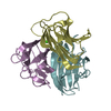
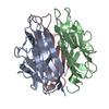
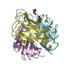
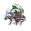
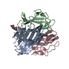
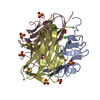
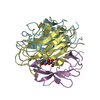
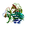
| Entry | Database: PDB / ID: 1o91 | ||||||
|---|---|---|---|---|---|---|---|
| Title | Crystal Structure of a Collagen VIII NC1 Domain Trimer | ||||||
 Components Components | COLLAGEN ALPHA 1(VIII) CHAIN | ||||||
 Keywords Keywords |  STRUCTURAL PROTEIN / COLLAGEN / C1Q_LIKE_DOMAIN / EXTRACELLULAR MATRIX / ADHESION / CONNECTIVE TISSUE STRUCTURAL PROTEIN / COLLAGEN / C1Q_LIKE_DOMAIN / EXTRACELLULAR MATRIX / ADHESION / CONNECTIVE TISSUE | ||||||
| Function / homology |  Function and homology information Function and homology informationCollagen biosynthesis and modifying enzymes / Collagen chain trimerization / Collagen degradation / Assembly of collagen fibrils and other multimeric structures / Integrin cell surface interactions / camera-type eye morphogenesis / collagen trimer / positive regulation of cell-substrate adhesion / extracellular matrix structural constituent / endodermal cell differentiation ...Collagen biosynthesis and modifying enzymes / Collagen chain trimerization / Collagen degradation / Assembly of collagen fibrils and other multimeric structures / Integrin cell surface interactions / camera-type eye morphogenesis / collagen trimer / positive regulation of cell-substrate adhesion / extracellular matrix structural constituent / endodermal cell differentiation / basement membrane / extracellular matrix organization / extracellular matrix / epithelial cell proliferation / angiogenesis / collagen-containing extracellular matrix / cell adhesion / extracellular spaceSimilarity search - Function | ||||||
| Biological species |  MUS MUSCULUS (house mouse) MUS MUSCULUS (house mouse) | ||||||
| Method | X-RAY DIFFRACTION / MOLECULAR REPLACEMENT / Resolution: 1.9 Å | ||||||
 Authors Authors | Kvansakul, M. / Bogin, O. / Hohenester, E. / Yayon, A. | ||||||
 Citation Citation | Journal: Matrix Biol. / Year: 2003 Title: Crystal Structure of the Collagen Alpha1(Viii) Nc1 Trimer. Authors: Kvansakul, M. / Bogin, O. / Hohenester, E. / Yayon, A. #1: Journal: Structure / Year: 2002Title: Insight Into Schmid Metaphyseal Chondrodysplasia from the Crystal Structure of the Collagen X Nc1 Domain Trimer. Authors: Bogin, O. / Kvansakul, M. / Rom, E. / Singer, J. / Yayon, A. / Hohenester, E. #2: Journal: Int. J. Biochem. Cell Biol. / Year: 1997 Title: Type Viii Collagen Authors: Shuttleworth, C.A. | ||||||
| History |
| ||||||
| Remark 700 | SHEET THE SHEET STRUCTURE OF THIS MOLECULE IS BIFURCATED. IN ORDER TO REPRESENT THIS FEATURE IN ... SHEET THE SHEET STRUCTURE OF THIS MOLECULE IS BIFURCATED. IN ORDER TO REPRESENT THIS FEATURE IN THE SHEET RECORDS BELOW, TWO SHEETS ARE DEFINED. |
- Structure visualization
| Structure viewer | Molecule: MolmilJmol/JSmol |
|---|
- Downloads & links
-Download
| PDBx/mmCIF format | 1o91.cif.gz | 98.5 KB | Display | PDBx/mmCIF format |
|---|---|---|---|---|
| PDB format | pdb1o91.ent.gz | 73.7 KB | Display | PDB format |
| PDBx/mmJSON format | 1o91.json.gz | Tree view | PDBx/mmJSON format | |
| Others |  Other downloads Other downloads |
-Validation report
| Arichive directory | https://data.pdbj.org/pub/pdb/validation_reports/o9/1o91ftp://data.pdbj.org/pub/pdb/validation_reports/o9/1o91 | HTTPS FTP |
|---|
-Related structure data
| Related structure data | 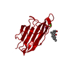 1gr3S S: Starting model for refinement |
|---|---|
| Similar structure data |


-Links
 PDBj
PDBj


- Assembly
| Deposited unit | 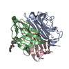
| ||||||||||||
|---|---|---|---|---|---|---|---|---|---|---|---|---|---|
| 1 |
| ||||||||||||
| Unit cell |
| ||||||||||||
| Noncrystallographic symmetry (NCS) | NCS oper:
|
-Components
| #1: Protein | Mass: 19734.426 Da / Num. of mol.: 3 / Fragment: NONHELICAL REGION (NC1), RESIDUES 613-743 Source method: isolated from a genetically manipulated source Source: (gene. exp.) MUS MUSCULUS (house mouse) / Plasmid: P89BLUESCRIPT / Production host:  ESCHERICHIA COLI (E. coli) / References: UniProt: Q00780 ESCHERICHIA COLI (E. coli) / References: UniProt: Q00780#2: Chemical | ChemComp-CPS / | CHAPS detergent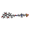  Mass: 614.877 Da / Num. of mol.: 1 / Source method: obtained synthetically / Formula: C32H58N2O7S / Comment: detergent*YM Mass: 614.877 Da / Num. of mol.: 1 / Source method: obtained synthetically / Formula: C32H58N2O7S / Comment: detergent*YM#3: Chemical | ChemComp-SO4 / | Sulfate  Mass: 96.063 Da / Num. of mol.: 1 / Source method: obtained synthetically / Formula: SO4 Mass: 96.063 Da / Num. of mol.: 1 / Source method: obtained synthetically / Formula: SO4#4: Water | ChemComp-HOH / | Water Mass: 18.015 Da / Num. of mol.: 268 / Source method: isolated from a natural source / Formula: H2O Mass: 18.015 Da / Num. of mol.: 268 / Source method: isolated from a natural source / Formula: H2O |
|---|
-Experimental details
-Experiment
| Experiment | Method: X-RAY DIFFRACTION / Number of used crystals: 1 |
|---|
- Sample preparation
| Crystal | Density Matthews: 2.1 Å3/Da / Density % sol: 43 % | |||||||||||||||||||||||||||||||||||||||||||||||||
|---|---|---|---|---|---|---|---|---|---|---|---|---|---|---|---|---|---|---|---|---|---|---|---|---|---|---|---|---|---|---|---|---|---|---|---|---|---|---|---|---|---|---|---|---|---|---|---|---|---|---|
| Crystal grow | pH: 7.5 Details: 1.7 M AMMONIUM SULFATE, 4 % (V/V)ISOPROPANOL, pH 7.50 | |||||||||||||||||||||||||||||||||||||||||||||||||
| Crystal grow | *PLUS pH: 9.5 / Method: vapor diffusion, hanging drop | |||||||||||||||||||||||||||||||||||||||||||||||||
| Components of the solutions | *PLUS
|
-Data collection
| Diffraction | Mean temperature: 100 K |
|---|---|
| Diffraction source | Source: ROTATING ANODE / Type: RIGAKU RUH3R / Wavelength: 1.5418 |
| Detector | Type: MARRESEARCH / Detector: IMAGE PLATE / Date: Jan 15, 2002 / Details: OSMIC MIRRORS |
| Radiation | Monochromator: GRAPHITE / Protocol: SINGLE WAVELENGTH / Monochromatic (M) / Laue (L): M / Scattering type: x-ray |
| Radiation wavelength | Wavelength: 1.5418 Å / Relative weight: 1 |
| Reflection | Resolution: 1.9→20 Å / Num. obs: 37444 / % possible obs: 98.9 % / Redundancy: 5.6 % / Biso Wilson estimate: 14.1 Å2 / Rmerge(I) obs: 0.087 / Net I/σ(I): 15.7 |
| Reflection shell | Resolution: 1.9→1.99 Å / Redundancy: 5.3 % / Rmerge(I) obs: 0.24 / Mean I/σ(I) obs: 6.7 / % possible all: 98.9 |
| Reflection | *PLUS Highest resolution: 1.9 Å / Lowest resolution: 20 Å / Num. obs: 37514 / Redundancy: 5.6 % / Rmerge(I) obs: 0.087 |
| Reflection shell | *PLUS % possible obs: 98.9 % / Redundancy: 5.3 % / Rmerge(I) obs: 0.24 |
- Processing
| Software |
| ||||||||||||||||||||||||||||||||||||||||||||||||||||||||||||||||||||||||||||||||
|---|---|---|---|---|---|---|---|---|---|---|---|---|---|---|---|---|---|---|---|---|---|---|---|---|---|---|---|---|---|---|---|---|---|---|---|---|---|---|---|---|---|---|---|---|---|---|---|---|---|---|---|---|---|---|---|---|---|---|---|---|---|---|---|---|---|---|---|---|---|---|---|---|---|---|---|---|---|---|---|---|---|
| Refinement | Method to determine structure: MOLECULAR REPLACEMENT Starting model: PDB ENTRY 1GR3 Resolution: 1.9→20 Å / Data cutoff high absF: 10000 / Isotropic thermal model: INDIVIDUALLY RESTRAINED / Cross valid method: THROUGHOUT / σ(F): 0
| ||||||||||||||||||||||||||||||||||||||||||||||||||||||||||||||||||||||||||||||||
| Solvent computation | Solvent model: FLAT / Bsol: 68 Å2 / ksol: 0.4 e/Å3 | ||||||||||||||||||||||||||||||||||||||||||||||||||||||||||||||||||||||||||||||||
| Refinement step | Cycle: LAST / Resolution: 1.9→20 Å
| ||||||||||||||||||||||||||||||||||||||||||||||||||||||||||||||||||||||||||||||||
| Refine LS restraints |
| ||||||||||||||||||||||||||||||||||||||||||||||||||||||||||||||||||||||||||||||||
| LS refinement shell | Resolution: 1.9→1.91 Å / Total num. of bins used: 50
| ||||||||||||||||||||||||||||||||||||||||||||||||||||||||||||||||||||||||||||||||
| Refinement | *PLUS Lowest resolution: 20 Å | ||||||||||||||||||||||||||||||||||||||||||||||||||||||||||||||||||||||||||||||||
| Solvent computation | *PLUS | ||||||||||||||||||||||||||||||||||||||||||||||||||||||||||||||||||||||||||||||||
| Displacement parameters | *PLUS |
