 Movie
Movie Controller


+ Open data

- Basic information
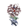
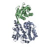
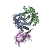
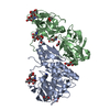
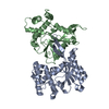
| Entry | Database: PDB / ID: 1nmu | |||||||||
|---|---|---|---|---|---|---|---|---|---|---|
| Title | MBP-L30 | |||||||||
 Components Components |
| |||||||||
 Keywords Keywords | SUGAR BINDING PROTEIN/RIBOSOME / structural flexibility /  ribosomal protein L30 / MBP-L30 fusion protein / SUGAR BINDING PROTEIN-RIBOSOME COMPLEX ribosomal protein L30 / MBP-L30 fusion protein / SUGAR BINDING PROTEIN-RIBOSOME COMPLEX | |||||||||
| Function / homology |  Function and homology information Function and homology informationpre-mRNA 5'-splice site binding / detection of maltose stimulus / maltose binding / maltose transport complex / maltose transport / maltodextrin transmembrane transport / SRP-dependent cotranslational protein targeting to membrane / GTP hydrolysis and joining of the 60S ribosomal subunit / Formation of a pool of free 40S subunits / Nonsense Mediated Decay (NMD) independent of the Exon Junction Complex (EJC) ...pre-mRNA 5'-splice site binding / detection of maltose stimulus / maltose binding / maltose transport complex / maltose transport / maltodextrin transmembrane transport / SRP-dependent cotranslational protein targeting to membrane / GTP hydrolysis and joining of the 60S ribosomal subunit / Formation of a pool of free 40S subunits / Nonsense Mediated Decay (NMD) independent of the Exon Junction Complex (EJC) / Nonsense Mediated Decay (NMD) enhanced by the Exon Junction Complex (EJC) / negative regulation of mRNA splicing, via spliceosome / L13a-mediated translational silencing of Ceruloplasmin expression / carbohydrate transmembrane transporter activity / ATP-binding cassette (ABC) transporter complex, substrate-binding subunit-containing / carbohydrate transport / ATP-binding cassette (ABC) transporter complex / cell chemotaxis / rRNA processing / outer membrane-bounded periplasmic space / cytoplasmic translation / cytosolic large ribosomal subunit / periplasmic space / structural constituent of ribosome / DNA damage response / RNA binding / membrane / cytosol / cytoplasmSimilarity search - Function | |||||||||
| Biological species |  Escherichia coli (E. coli) Escherichia coli (E. coli) Saccharomyces cerevisiae (brewer's yeast) Saccharomyces cerevisiae (brewer's yeast) | |||||||||
| Method | X-RAY DIFFRACTION / SYNCHROTRON / MOLECULAR REPLACEMENT / Resolution: 2.31 Å | |||||||||
 Authors Authors | Chao, J.A. / Prasad, G.S. / White, S.A. / Stout, C.D. / Williamson, J.R. | |||||||||
 Citation Citation | Journal: J.Mol.Biol. / Year: 2003 Title: Inherent Protein Structural Flexibility at the RNA-binding Interface of L30e Authors: Chao, J.A. / Prasad, G.S. / White, S.A. / Stout, C.D. / Williamson, J.R. | |||||||||
| History |
|
- Structure visualization
| Structure viewer | Molecule: MolmilJmol/JSmol |
|---|
- Downloads & links
-Download
| PDBx/mmCIF format | 1nmu.cif.gz | 190.6 KB | Display | PDBx/mmCIF format |
|---|---|---|---|---|
| PDB format | pdb1nmu.ent.gz | 155.4 KB | Display | PDB format |
| PDBx/mmJSON format | 1nmu.json.gz | Tree view | PDBx/mmJSON format | |
| Others |  Other downloads Other downloads |
-Validation report
| Arichive directory | https://data.pdbj.org/pub/pdb/validation_reports/nm/1nmuftp://data.pdbj.org/pub/pdb/validation_reports/nm/1nmu | HTTPS FTP |
|---|
-Related structure data
| Similar structure data |
|---|


-Links
 PDBj
PDBj


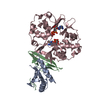
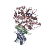
- Assembly
| Deposited unit | 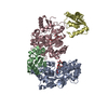
| ||||||||
|---|---|---|---|---|---|---|---|---|---|
| 1 | 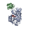
| ||||||||
| 2 | 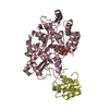
| ||||||||
| Unit cell |
|
-Components
| #1: Protein | Mass: 41841.266 Da / Num. of mol.: 2 Source method: isolated from a genetically manipulated source Source: (gene. exp.) Escherichia coli (E. coli) / Production host: Escherichia coli (E. coli) / References: UniProt: P02928, UniProt: P0AEX9*PLUS#2: Protein | / YL32 / RP73Mass: 11299.168 Da / Num. of mol.: 2 Source method: isolated from a genetically manipulated source Source: (gene. exp.) Saccharomyces cerevisiae (brewer's yeast)Production host: Saccharomyces cerevisiae (brewer's yeast) / References: UniProt: P14120#3: Polysaccharide | 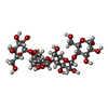  , Oligosaccharide / Class: Substrate analog / Mass: 666.578 Da / Num. of mol.: 2 , Oligosaccharide / Class: Substrate analog / Mass: 666.578 Da / Num. of mol.: 2Source method: isolated from a genetically manipulated source Details: oligosaccharide / References: alpha-maltotetraose #4: Water | ChemComp-HOH / | Water Mass: 18.015 Da / Num. of mol.: 232 / Source method: isolated from a natural source / Formula: H2O Mass: 18.015 Da / Num. of mol.: 232 / Source method: isolated from a natural source / Formula: H2O |
|---|
-Experimental details
-Experiment
| Experiment | Method: X-RAY DIFFRACTION / Number of used crystals: 1 |
|---|
- Sample preparation
| Crystal | Density Matthews: 3.41 Å3/Da / Density % sol: 63.92 % | ||||||||||||||||||||||||||||||||||||||||||
|---|---|---|---|---|---|---|---|---|---|---|---|---|---|---|---|---|---|---|---|---|---|---|---|---|---|---|---|---|---|---|---|---|---|---|---|---|---|---|---|---|---|---|---|
| Crystal grow | Temperature: 295 K / Method: vapor diffusion, hanging drop / pH: 7 Details: Sodium Citrate, Tris, Sodium Chloride, pH 7, VAPOR DIFFUSION, HANGING DROP, temperature 295K | ||||||||||||||||||||||||||||||||||||||||||
| Crystal grow | *PLUS Temperature: 22 ℃ / pH: 6.2 | ||||||||||||||||||||||||||||||||||||||||||
| Components of the solutions | *PLUS
|
-Data collection
| Diffraction | Mean temperature: 93 K |
|---|---|
| Diffraction source | Source: SYNCHROTRON / Site: SSRL  / Beamline: BL9-2 / Wavelength: 0.9795 Å / Beamline: BL9-2 / Wavelength: 0.9795 Å |
| Detector | Type: ADSC QUANTUM 4 / Detector: CCD / Date: Nov 16, 2000 / Details: double crystal monochromater |
| Radiation | Monochromator: graphite / Protocol: SINGLE WAVELENGTH / Monochromatic (M) / Laue (L): M / Scattering type: x-ray |
| Radiation wavelength | Wavelength: 0.9795 Å / Relative weight: 1 |
| Reflection | Resolution: 2.31→18 Å / Num. all: 65857 / Num. obs: 65330 / % possible obs: 99.2 % / Observed criterion σ(F): 2 / Observed criterion σ(I): 2 |
| Reflection shell | Resolution: 2.31→2.38 Å / % possible all: 99.8 |
| Reflection | *PLUS Lowest resolution: 18 Å / Num. measured all: 708695 / Rmerge(I) obs: 0.086 |
| Reflection shell | *PLUS % possible obs: 99.8 % / Rmerge(I) obs: 0.359 |
- Processing
| Software |
| ||||||||||||||||||||
|---|---|---|---|---|---|---|---|---|---|---|---|---|---|---|---|---|---|---|---|---|---|
| Refinement | Method to determine structure: MOLECULAR REPLACEMENT / Resolution: 2.31→18 Å / Cross valid method: THROUGHOUT / σ(F): 0 / Stereochemistry target values: Engh & Huber
| ||||||||||||||||||||
| Refinement step | Cycle: LAST / Resolution: 2.31→18 Å
| ||||||||||||||||||||
| Refine LS restraints |
| ||||||||||||||||||||
| Refinement | *PLUS Lowest resolution: 18 Å / Rfactor Rfree: 0.2544 / Rfactor Rwork: 0.2225 | ||||||||||||||||||||
| Solvent computation | *PLUS | ||||||||||||||||||||
| Displacement parameters | *PLUS | ||||||||||||||||||||
| Refine LS restraints | *PLUS
|
