 Movie
Movie Controller

[English] 日本語
 Yorodumi
- PDB-5upl: CDC42 binds PAK4 via an extended GTPase-effector inteface - 2 pep... -
+ Open data

- Basic information
| Entry | Database: PDB / ID: 5upl | ||||||
|---|---|---|---|---|---|---|---|
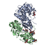
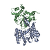
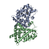
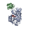
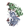
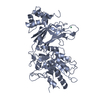
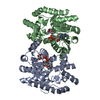
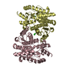
| Title | CDC42 binds PAK4 via an extended GTPase-effector inteface - 2 peptide: PAK4FL, CDC42 - UNREFINED | ||||||
 Components Components |
| ||||||
 Keywords Keywords |  TRANSFERASE / GTPase / Kinase / CRIB TRANSFERASE / GTPase / Kinase / CRIB | ||||||
| Function / homology |  Function and homology information Function and homology informationGBD domain binding / submandibular salivary gland formation / actin filament branching / Golgi transport complex / positive regulation of pinocytosis / modification of synaptic structure / endothelin receptor signaling pathway involved in heart process / Cdc42 protein signal transduction / cardiac neural crest cell migration involved in outflow tract morphogenesis / positive regulation of synapse structural plasticity ...GBD domain binding / submandibular salivary gland formation / actin filament branching / Golgi transport complex / positive regulation of pinocytosis / modification of synaptic structure / endothelin receptor signaling pathway involved in heart process / Cdc42 protein signal transduction / cardiac neural crest cell migration involved in outflow tract morphogenesis / positive regulation of synapse structural plasticity / dendritic cell migration / storage vacuole / positive regulation of epithelial cell proliferation involved in lung morphogenesis / apolipoprotein A-I receptor binding / neuron fate determination / modulation by host of viral process / GTP-dependent protein binding / organelle transport along microtubule / regulation of attachment of spindle microtubules to kinetochore / positive regulation of pseudopodium assembly / Inactivation of CDC42 and RAC1 / cardiac conduction system development / dendritic spine development / cadherin binding involved in cell-cell adhesion / regulation of filopodium assembly / establishment of Golgi localization / leading edge membrane / neuropilin signaling pathway / positive regulation of intracellular protein transport / cell junction assembly / filopodium assembly / establishment of epithelial cell apical/basal polarity / regulation of modification of postsynaptic structure / mitogen-activated protein kinase kinase kinase binding / dendritic spine morphogenesis / thioesterase binding / embryonic heart tube development / regulation of stress fiber assembly / RHO GTPases activate KTN1 / Activation of RAC1 / regulation of lamellipodium assembly / nuclear migration / DCC mediated attractive signaling / adherens junction organization / sprouting angiogenesis / Wnt signaling pathway, planar cell polarity pathway / CD28 dependent Vav1 pathway / regulation of postsynapse organization / positive regulation of filopodium assembly / regulation of mitotic nuclear division / phagocytosis, engulfment / RHOV GTPase cycle / establishment or maintenance of cell polarity / heart contraction / Myogenesis / RHOJ GTPase cycle / Golgi organization / RHOQ GTPase cycle / positive regulation of cytokinesis / RHO GTPases activate PAKs / regulation of MAPK cascade / RHOH GTPase cycle / CDC42 GTPase cycle / RHOU GTPase cycle / macrophage differentiation / cellular response to organic cyclic compound / RHOG GTPase cycle / RHO GTPases Activate WASPs and WAVEs / RAC2 GTPase cycle / RHO GTPases activate IQGAPs / RAC3 GTPase cycle / spindle midzone / negative regulation of protein-containing complex assembly / positive regulation of lamellipodium assembly / negative regulation of endothelial cell apoptotic process / positive regulation of substrate adhesion-dependent cell spreading / phagocytic vesicle / positive regulation of stress fiber assembly / GPVI-mediated activation cascade / cytoskeleton organization / RAC1 GTPase cycle / EPHB-mediated forward signaling / substantia nigra development / Gene and protein expression by JAK-STAT signaling after Interleukin-12 stimulation / small monomeric GTPase / G protein activity / secretory granule / positive regulation of DNA replication / filopodium / integrin-mediated signaling pathway / RHO GTPases Activate Formins / actin filament organization / regulation of cell growth / regulation of actin cytoskeleton organization / FCGR3A-mediated phagocytosis / EGFR downregulation / positive regulation of JNK cascade / adherens junction / MAPK6/MAPK4 signaling / Schaffer collateral - CA1 synapseSimilarity search - Function | ||||||
| Biological species |  Homo sapiens (human) Homo sapiens (human) | ||||||
| Method | X-RAY DIFFRACTION / SYNCHROTRON / MOLECULAR REPLACEMENT / Resolution: 3.003 Å | ||||||
 Authors Authors | Ha, B.H. / Boggon, T.J. | ||||||
| Funding support |  United States, 1items United States, 1items
| ||||||
 Citation Citation | Journal: Proc. Natl. Acad. Sci. U.S.A. / Year: 2018 Title: CDC42 binds PAK4 via an extended GTPase-effector interface. Authors: Ha, B.H. / Boggon, T.J. | ||||||
| History |
|
- Structure visualization
| Structure viewer | Molecule: MolmilJmol/JSmol |
|---|
- Downloads & links
-Download
| PDBx/mmCIF format | 5upl.cif.gz | 104.8 KB | Display | PDBx/mmCIF format |
|---|---|---|---|---|
| PDB format | pdb5upl.ent.gz | 75.7 KB | Display | PDB format |
| PDBx/mmJSON format | 5upl.json.gz | Tree view | PDBx/mmJSON format | |
| Others |  Other downloads Other downloads |
-Validation report
| Arichive directory | https://data.pdbj.org/pub/pdb/validation_reports/up/5uplftp://data.pdbj.org/pub/pdb/validation_reports/up/5upl | HTTPS FTP |
|---|
-Related structure data
| Related structure data |  5upkC 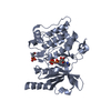 4fieS C: citing same article ( S: Starting model for refinement |
|---|---|
| Similar structure data |


-Links
 PDBj
PDBj


- Assembly
| Deposited unit | 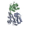
| ||||||||
|---|---|---|---|---|---|---|---|---|---|
| 1 |
| ||||||||
| Unit cell |
|
-Components
| #1: Protein | Mass: 51099.684 Da / Num. of mol.: 1 / Fragment: UNP residues 2-426 / Mutation: S474SEP Source method: isolated from a genetically manipulated source Details: Ser474 is phosphorylated (SEP) / Source: (gene. exp.) Homo sapiens (human) / Gene: PAK4, KIAA1142 / Variant: isoform2 / Plasmid: modified pET28a / Production host:  Escherichia coli (E. coli) / Strain (production host): BL21(DE3)RILP Escherichia coli (E. coli) / Strain (production host): BL21(DE3)RILPReferences: UniProt: O96013, non-specific serine/threonine protein kinase |
|---|---|
| #2: Protein | Mass: 20716.744 Da / Num. of mol.: 1 / Fragment: UNP residues 1-177 Source method: isolated from a genetically manipulated source Source: (gene. exp.) Homo sapiens (human) / Gene: CDC42 / Plasmid: pET22b / Production host: Escherichia coli (E. coli) / Strain (production host): BL21(DE3)RILP / References: UniProt: P60953 |
-Experimental details
-Experiment
| Experiment | Method: X-RAY DIFFRACTION / Number of used crystals: 1 |
|---|
- Sample preparation
| Crystal | Density Matthews: 2.5 Å3/Da / Density % sol: 50.88 % |
|---|---|
| Crystal grow | Temperature: 295 K / Method: vapor diffusion, hanging drop / pH: 8.5 / Details: 0.1M Tris-HCl pH 8.5, 50mM Na2SO4, 6% PEG6000 / PH range: 8.0-9.0 / Temp details: R/T |
-Data collection
| Diffraction | Mean temperature: 100 K |
|---|---|
| Diffraction source | Source: SYNCHROTRON / Site: APS / Beamline: 24-ID-E / Wavelength: 0.97922 Å |
| Detector | Type: ADSC QUANTUM 315 / Detector: CCD / Date: Dec 9, 2013 |
| Radiation | Monochromator: Si (111) / Protocol: SINGLE WAVELENGTH / Monochromatic (M) / Laue (L): M / Scattering type: x-ray |
| Radiation wavelength | Wavelength: 0.97922 Å / Relative weight: 1 |
| Reflection | Resolution: 3→43.637 Å / Num. obs: 14433 / % possible obs: 99.9 % / Redundancy: 18 % / CC1/2: 0.588 / Rmerge(I) obs: 0.117 / Rpim(I) all: 0.034 / Rsym value: 0.117 / Χ2: 1.187 / Net I/σ(I): 20.3 |
| Reflection shell | Resolution: 3→3.11 Å / Mean I/σ(I) obs: 1.6 / CC1/2: 0.588 / Rpim(I) all: 0.707 / Χ2: 1.608 / % possible all: 100 |
- Processing
| Software |
| ||||||||||||||||||||||||||||||||||||||||||
|---|---|---|---|---|---|---|---|---|---|---|---|---|---|---|---|---|---|---|---|---|---|---|---|---|---|---|---|---|---|---|---|---|---|---|---|---|---|---|---|---|---|---|---|
| Refinement | Method to determine structure: MOLECULAR REPLACEMENT Starting model: 4fie Resolution: 3.003→43.637 Å / SU ML: 0.57 / Cross valid method: NONE / σ(F): 1.33 / Phase error: 34.46 / Stereochemistry target values: ML
| ||||||||||||||||||||||||||||||||||||||||||
| Solvent computation | Shrinkage radii: 0.9 Å / VDW probe radii: 1.11 Å / Solvent model: FLAT BULK SOLVENT MODEL | ||||||||||||||||||||||||||||||||||||||||||
| Refinement step | Cycle: LAST / Resolution: 3.003→43.637 Å
| ||||||||||||||||||||||||||||||||||||||||||
| Refine LS restraints |
| ||||||||||||||||||||||||||||||||||||||||||
| LS refinement shell |
|
