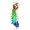
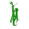
Mitochondrial protein import protein MAS5Mitochondrion
Seven residue peptide
Keywords
PROTEIN TRANSPORT / beta-strands / chaperone / heat shock / mitochondrion
Function / homology
Function and homology information
tRNA import into nucleus / TRC complex / HSP90 chaperone cycle for steroid hormone receptors (SHR) in the presence of ligand / response to oxygen levels / protein targeting to ER / protein targeting to mitochondrion / 'de novo' protein folding / ATPase activator activity / chaperone-mediated protein complex assembly / : ...tRNA import into nucleus / TRC complex / HSP90 chaperone cycle for steroid hormone receptors (SHR) in the presence of ligand / response to oxygen levels / protein targeting to ER / protein targeting to mitochondrion / 'de novo' protein folding / ATPase activator activity / chaperone-mediated protein complex assembly / : / Hsp70 protein binding / transcription repressor complex / unfolded protein binding / protein transport / response to heat / ubiquitin-dependent protein catabolic process / protein-folding chaperone binding / protein refolding / negative regulation of DNA-templated transcription / perinuclear region of cytoplasm / ATP binding / metal ion binding / nucleus / cytosol / cytoplasm Similarity search - Function
In the structure databanks used in Yorodumi, some data are registered as the other names, "COVID-19 virus" and "2019-nCoV". Here are the details of the virus and the list of structure data.
Jan 31, 2019. EMDB accession codes are about to change! (news from PDBe EMDB page)
EMDB accession codes are about to change! (news from PDBe EMDB page)
The allocation of 4 digits for EMDB accession codes will soon come to an end. Whilst these codes will remain in use, new EMDB accession codes will include an additional digit and will expand incrementally as the available range of codes is exhausted. The current 4-digit format prefixed with “EMD-” (i.e. EMD-XXXX) will advance to a 5-digit format (i.e. EMD-XXXXX), and so on. It is currently estimated that the 4-digit codes will be depleted around Spring 2019, at which point the 5-digit format will come into force.
The EM Navigator/Yorodumi systems omit the EMD- prefix.
Related info.:Q: What is EMD? / ID/Accession-code notation in Yorodumi/EM Navigator
Yorodumi is a browser for structure data from EMDB, PDB, SASBDB, etc.
This page is also the successor to EM Navigator detail page, and also detail information page/front-end page for Omokage search.
The word "yorodu" (or yorozu) is an old Japanese word meaning "ten thousand". "mi" (miru) is to see.
Related info.:EMDB / PDB / SASBDB / Comparison of 3 databanks / Yorodumi Search / Aug 31, 2016. New EM Navigator & Yorodumi / Yorodumi Papers / Jmol/JSmol / Function and homology information / Changes in new EM Navigator and Yorodumi
 Movie
Movie Controller


 Open data
 Basic information
 Components
 Keywords
 PROTEIN TRANSPORT / beta-strands /
  Function and homology information
 Authors
 Citation
 Structure visualization
 Downloads & links
 Other downloads
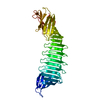


 PDBj
PDBj


 Assembly


 Mass: 65.409 Da / Num. of mol.: 2 / Source method: obtained synthetically / Formula: Zn
 Mass: 18.015 Da / Num. of mol.: 147 / Source method: isolated from a natural source / Formula: H2O
 Sample preparation
 Processing