 Movie
Movie Controller

[English] 日本語
 Yorodumi
- PDB-5y4f: Crystal Structure of AnkB Ankyrin Repeats R13-24 in complex with ... -
+ Open data

- Basic information
| Entry | Database: PDB / ID: 5y4f | ||||||
|---|---|---|---|---|---|---|---|
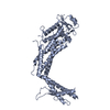
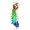
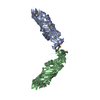
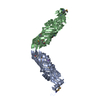
| Title | Crystal Structure of AnkB Ankyrin Repeats R13-24 in complex with autoinhibition segment AI-c | ||||||
 Components Components | Ankyrin-2 | ||||||
 Keywords Keywords | PROTEIN BINDING / ANK REPEAT / PROTEIN-PROTEIN INTERACTION / STRUCTURAL PROTEIN / AUTO-INHIBITION | ||||||
| Function / homology |  Function and homology information Function and homology informationprotein localization to T-tubule / atrial cardiac muscle cell to AV node cell communication / SA node cell to atrial cardiac muscle cell communication / regulation of calcium ion transmembrane transporter activity / positive regulation of calcium ion transmembrane transporter activity / protein localization to M-band / positive regulation of potassium ion transmembrane transporter activity / T-tubule organization / SA node cell action potential / membrane depolarization during SA node cell action potential ...protein localization to T-tubule / atrial cardiac muscle cell to AV node cell communication / SA node cell to atrial cardiac muscle cell communication / regulation of calcium ion transmembrane transporter activity / positive regulation of calcium ion transmembrane transporter activity / protein localization to M-band / positive regulation of potassium ion transmembrane transporter activity / T-tubule organization / SA node cell action potential / membrane depolarization during SA node cell action potential / protein localization to organelle / paranodal junction assembly / phosphorylation-dependent protein binding / regulation of SA node cell action potential / regulation of atrial cardiac muscle cell action potential / protein localization to endoplasmic reticulum / positive regulation of cation channel activity / atrial cardiac muscle cell action potential / sarcoplasmic reticulum calcium ion transport / cytoskeletal anchor activity / atrial septum development / positive regulation of potassium ion transport / ventricular cardiac muscle cell action potential / costamere / positive regulation of calcium ion transport / regulation of release of sequestered calcium ion into cytosol / response to methylmercury / regulation of ventricular cardiac muscle cell membrane repolarization / M band / regulation of cardiac muscle cell contraction / protein localization to cell surface / Interaction between L1 and Ankyrins / regulation of cardiac muscle contraction by calcium ion signaling / A band / spectrin binding / regulation of heart rate by cardiac conduction / regulation of calcium ion transport / intercalated disc / regulation of cardiac muscle contraction / regulation of cardiac muscle contraction by regulation of the release of sequestered calcium ion / COPI-mediated anterograde transport / T-tubule / regulation of heart rate / protein localization to plasma membrane / regulation of protein stability / protein localization / recycling endosome / structural constituent of cytoskeleton / sarcolemma / Z disc / intracellular calcium ion homeostasis / endocytosis / protein transport / protein-macromolecule adaptor activity / ATPase binding / basolateral plasma membrane / postsynaptic membrane / transmembrane transporter binding / lysosome / early endosome / cytoskeleton / protein stabilization / neuron projection / apical plasma membrane / positive regulation of gene expression / protein kinase binding / enzyme binding / mitochondrion / plasma membrane / cytosolSimilarity search - Function | ||||||
| Biological species |  Homo sapiens (human) Homo sapiens (human) | ||||||
| Method | X-RAY DIFFRACTION / SYNCHROTRON / MOLECULAR REPLACEMENT / Resolution: 1.953 Å | ||||||
 Authors Authors | Chen, K. / Li, J. / Wang, C. / Wei, Z. / Zhang, M. | ||||||
 Citation Citation | Journal: Elife / Year: 2017 Title: Autoinhibition of ankyrin-B/G membrane target bindings by intrinsically disordered segments from the tail regions. Authors: Chen, K. / Li, J. / Wang, C. / Wei, Z. / Zhang, M. | ||||||
| History |
|
- Structure visualization
| Structure viewer | Molecule: MolmilJmol/JSmol |
|---|
- Downloads & links
-Download
| PDBx/mmCIF format | 5y4f.cif.gz | 318.1 KB | Display | PDBx/mmCIF format |
|---|---|---|---|---|
| PDB format | pdb5y4f.ent.gz | 256.8 KB | Display | PDB format |
| PDBx/mmJSON format | 5y4f.json.gz | Tree view | PDBx/mmJSON format | |
| Others |  Other downloads Other downloads |
-Validation report
| Arichive directory | https://data.pdbj.org/pub/pdb/validation_reports/y4/5y4fftp://data.pdbj.org/pub/pdb/validation_reports/y4/5y4f | HTTPS FTP |
|---|
-Related structure data
| Related structure data |  5y4dC  5y4eC  4rlvS S: Starting model for refinement C: citing same article ( |
|---|---|
| Similar structure data |


-Links
 PDBj
PDBj


- Assembly
| Deposited unit | 
| ||||||||
|---|---|---|---|---|---|---|---|---|---|
| 1 | 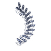
| ||||||||
| 2 | 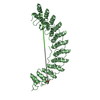
| ||||||||
| Unit cell |
|
-Components
| #1: Protein | / ANK-2 / Ankyrin-B / Brain ankyrin / Non-erythroid ankyrin Mass: 47356.727 Da / Num. of mol.: 2 / Fragment: UNP RESIDUES 430-873 Source method: isolated from a genetically manipulated source Source: (gene. exp.) Homo sapiens (human) / Gene: ANK2 / Production host:  Escherichia coli (E. coli) / References: UniProt: Q01484 Escherichia coli (E. coli) / References: UniProt: Q01484#2: Chemical | ChemComp-CA / |   Mass: 40.078 Da / Num. of mol.: 1 / Source method: obtained synthetically / Formula: Ca Mass: 40.078 Da / Num. of mol.: 1 / Source method: obtained synthetically / Formula: Ca#3: Chemical | ChemComp-ACT / | Acetate  Mass: 59.044 Da / Num. of mol.: 1 / Source method: obtained synthetically / Formula: C2H3O2 Mass: 59.044 Da / Num. of mol.: 1 / Source method: obtained synthetically / Formula: C2H3O2#4: Water | ChemComp-HOH / | Water Mass: 18.015 Da / Num. of mol.: 274 / Source method: isolated from a natural source / Formula: H2O Mass: 18.015 Da / Num. of mol.: 274 / Source method: isolated from a natural source / Formula: H2O |
|---|
-Experimental details
-Experiment
| Experiment | Method: X-RAY DIFFRACTION / Number of used crystals: 1 |
|---|
- Sample preparation
| Crystal | Density Matthews: 2.55 Å3/Da / Density % sol: 51.69 % |
|---|---|
| Crystal grow | Temperature: 289 K / Method: vapor diffusion, hanging drop / pH: 7.5 / Details: 0.2 M CaCl2, 0.1 M HEPES (pH 7.5), 28% v/v PEG 400 |
-Data collection
| Diffraction | Mean temperature: 100 K | |||||||||||||||||||||||||||||||||||||||||||||||||||||||||||||||||||||||||||||||||||||||||||||||||||||||||||||||||||||||||||||||||||||||||||||||||||
|---|---|---|---|---|---|---|---|---|---|---|---|---|---|---|---|---|---|---|---|---|---|---|---|---|---|---|---|---|---|---|---|---|---|---|---|---|---|---|---|---|---|---|---|---|---|---|---|---|---|---|---|---|---|---|---|---|---|---|---|---|---|---|---|---|---|---|---|---|---|---|---|---|---|---|---|---|---|---|---|---|---|---|---|---|---|---|---|---|---|---|---|---|---|---|---|---|---|---|---|---|---|---|---|---|---|---|---|---|---|---|---|---|---|---|---|---|---|---|---|---|---|---|---|---|---|---|---|---|---|---|---|---|---|---|---|---|---|---|---|---|---|---|---|---|---|---|---|---|
| Diffraction source | Source: SYNCHROTRON / Site: SSRF  / Beamline: BL17U1 / Wavelength: 0.979 Å / Beamline: BL17U1 / Wavelength: 0.979 Å | |||||||||||||||||||||||||||||||||||||||||||||||||||||||||||||||||||||||||||||||||||||||||||||||||||||||||||||||||||||||||||||||||||||||||||||||||||
| Detector | Type: ADSC QUANTUM 315r / Detector: CCD / Date: Sep 14, 2013 | |||||||||||||||||||||||||||||||||||||||||||||||||||||||||||||||||||||||||||||||||||||||||||||||||||||||||||||||||||||||||||||||||||||||||||||||||||
| Radiation | Protocol: SINGLE WAVELENGTH / Monochromatic (M) / Laue (L): M / Scattering type: x-ray | |||||||||||||||||||||||||||||||||||||||||||||||||||||||||||||||||||||||||||||||||||||||||||||||||||||||||||||||||||||||||||||||||||||||||||||||||||
| Radiation wavelength | Wavelength: 0.979 Å / Relative weight: 1 | |||||||||||||||||||||||||||||||||||||||||||||||||||||||||||||||||||||||||||||||||||||||||||||||||||||||||||||||||||||||||||||||||||||||||||||||||||
| Reflection | Resolution: 1.95→50 Å / Num. obs: 68019 / % possible obs: 94.4 % / Redundancy: 3.1 % / Biso Wilson estimate: 27.19 Å2 / Rmerge(I) obs: 0.082 / Χ2: 2.497 / Net I/σ(I): 13.2 | |||||||||||||||||||||||||||||||||||||||||||||||||||||||||||||||||||||||||||||||||||||||||||||||||||||||||||||||||||||||||||||||||||||||||||||||||||
| Reflection shell |
|
- Processing
| Software |
| |||||||||||||||||||||||||||||||||||||||||||||||||||||||||||||||||||||||||||||||||||||||||||||||||||||||||||||||||||||||||||||||||||||||||||||||||||||||||||||||||||||||||||||||
|---|---|---|---|---|---|---|---|---|---|---|---|---|---|---|---|---|---|---|---|---|---|---|---|---|---|---|---|---|---|---|---|---|---|---|---|---|---|---|---|---|---|---|---|---|---|---|---|---|---|---|---|---|---|---|---|---|---|---|---|---|---|---|---|---|---|---|---|---|---|---|---|---|---|---|---|---|---|---|---|---|---|---|---|---|---|---|---|---|---|---|---|---|---|---|---|---|---|---|---|---|---|---|---|---|---|---|---|---|---|---|---|---|---|---|---|---|---|---|---|---|---|---|---|---|---|---|---|---|---|---|---|---|---|---|---|---|---|---|---|---|---|---|---|---|---|---|---|---|---|---|---|---|---|---|---|---|---|---|---|---|---|---|---|---|---|---|---|---|---|---|---|---|---|---|---|---|
| Refinement | Method to determine structure: MOLECULAR REPLACEMENT Starting model: 4RLV Resolution: 1.953→38.161 Å / SU ML: 0.18 / Cross valid method: FREE R-VALUE / σ(F): 1.34 / Phase error: 22.04
| |||||||||||||||||||||||||||||||||||||||||||||||||||||||||||||||||||||||||||||||||||||||||||||||||||||||||||||||||||||||||||||||||||||||||||||||||||||||||||||||||||||||||||||||
| Solvent computation | Shrinkage radii: 0.9 Å / VDW probe radii: 1.11 Å | |||||||||||||||||||||||||||||||||||||||||||||||||||||||||||||||||||||||||||||||||||||||||||||||||||||||||||||||||||||||||||||||||||||||||||||||||||||||||||||||||||||||||||||||
| Displacement parameters | Biso max: 101.23 Å2 / Biso mean: 30.9267 Å2 / Biso min: 12.41 Å2 | |||||||||||||||||||||||||||||||||||||||||||||||||||||||||||||||||||||||||||||||||||||||||||||||||||||||||||||||||||||||||||||||||||||||||||||||||||||||||||||||||||||||||||||||
| Refinement step | Cycle: final / Resolution: 1.953→38.161 Å
| |||||||||||||||||||||||||||||||||||||||||||||||||||||||||||||||||||||||||||||||||||||||||||||||||||||||||||||||||||||||||||||||||||||||||||||||||||||||||||||||||||||||||||||||
| Refine LS restraints |
| |||||||||||||||||||||||||||||||||||||||||||||||||||||||||||||||||||||||||||||||||||||||||||||||||||||||||||||||||||||||||||||||||||||||||||||||||||||||||||||||||||||||||||||||
| LS refinement shell | Refine-ID: X-RAY DIFFRACTION / Rfactor Rfree error: 0 / Total num. of bins used: 24
| |||||||||||||||||||||||||||||||||||||||||||||||||||||||||||||||||||||||||||||||||||||||||||||||||||||||||||||||||||||||||||||||||||||||||||||||||||||||||||||||||||||||||||||||
| Refinement TLS params. | Method: refined / Refine-ID: X-RAY DIFFRACTION
| |||||||||||||||||||||||||||||||||||||||||||||||||||||||||||||||||||||||||||||||||||||||||||||||||||||||||||||||||||||||||||||||||||||||||||||||||||||||||||||||||||||||||||||||
| Refinement TLS group |
|
