 Movie
Movie Controller


+ Open data

- Basic information
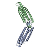
| Entry | Database: PDB / ID: 6dv1 | ||||||
|---|---|---|---|---|---|---|---|
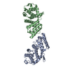
| Title | Crystal structure of the alpha-E-catenin actin-binding domain | ||||||
 Components Components | Catenin alpha-1 | ||||||
 Keywords Keywords | CELL ADHESION / five-helix bundle / F-actin-binding / mechanosensor | ||||||
| Function / homology |  Function and homology information Function and homology informationnegative regulation of integrin-mediated signaling pathway / VEGFR2 mediated vascular permeability / RHO GTPases activate IQGAPs / Adherens junctions interactions / gap junction assembly / epithelial cell-cell adhesion / zonula adherens / gamma-catenin binding / cellular response to indole-3-methanol / vinculin binding ...negative regulation of integrin-mediated signaling pathway / VEGFR2 mediated vascular permeability / RHO GTPases activate IQGAPs / Adherens junctions interactions / gap junction assembly / epithelial cell-cell adhesion / zonula adherens / gamma-catenin binding / cellular response to indole-3-methanol / vinculin binding / flotillin complex / negative regulation of cell motility / Myogenesis / apical junction assembly / positive regulation of extrinsic apoptotic signaling pathway in absence of ligand / positive regulation of smoothened signaling pathway / catenin complex / negative regulation of protein localization to nucleus / axon regeneration / negative regulation of neuroblast proliferation / smoothened signaling pathway / establishment or maintenance of cell polarity / odontogenesis of dentin-containing tooth / neuroblast proliferation / negative regulation of extrinsic apoptotic signaling pathway in absence of ligand / intercalated disc / ovarian follicle development / extrinsic apoptotic signaling pathway in absence of ligand / acrosomal vesicle / cell motility / integrin-mediated signaling pathway / adherens junction / protein localization / cell-cell adhesion / beta-catenin binding / response to estrogen / male gonad development / cell migration / actin filament binding / cell-cell junction / actin cytoskeleton / cell junction / lamellipodium / regulation of cell population proliferation / cadherin binding / intracellular membrane-bounded organelle / apoptotic process / protein-containing complex binding / negative regulation of apoptotic process / structural molecule activity / identical protein binding / plasma membraneSimilarity search - Function | ||||||
| Biological species |  Mus musculus (house mouse) Mus musculus (house mouse) | ||||||
| Method | X-RAY DIFFRACTION / SYNCHROTRON / SAD / Resolution: 2.2 Å | ||||||
 Authors Authors | Ishiyama, N. / Ikura, M. | ||||||
| Funding support |  Canada, 1items Canada, 1items
| ||||||
 Citation Citation | Journal: Nat Commun / Year: 2018 Title: Force-dependent allostery of the alpha-catenin actin-binding domain controls adherens junction dynamics and functions. Authors: Ishiyama, N. / Sarpal, R. / Wood, M.N. / Barrick, S.K. / Nishikawa, T. / Hayashi, H. / Kobb, A.B. / Flozak, A.S. / Yemelyanov, A. / Fernandez-Gonzalez, R. / Yonemura, S. / Leckband, D.E. / ...Authors: Ishiyama, N. / Sarpal, R. / Wood, M.N. / Barrick, S.K. / Nishikawa, T. / Hayashi, H. / Kobb, A.B. / Flozak, A.S. / Yemelyanov, A. / Fernandez-Gonzalez, R. / Yonemura, S. / Leckband, D.E. / Gottardi, C.J. / Tepass, U. / Ikura, M. | ||||||
| History |
|
- Structure visualization
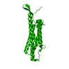
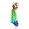
| Structure viewer | Molecule: MolmilJmol/JSmol |
|---|
- Downloads & links
-Download
| PDBx/mmCIF format | 6dv1.cif.gz | 108.6 KB | Display | PDBx/mmCIF format |
|---|---|---|---|---|
| PDB format | pdb6dv1.ent.gz | 66.8 KB | Display | PDB format |
| PDBx/mmJSON format | 6dv1.json.gz | Tree view | PDBx/mmJSON format | |
| Others |  Other downloads Other downloads |
-Validation report
| Arichive directory | https://data.pdbj.org/pub/pdb/validation_reports/dv/6dv1ftp://data.pdbj.org/pub/pdb/validation_reports/dv/6dv1 | HTTPS FTP |
|---|
-Related structure data


-Links
 PDBj
PDBj


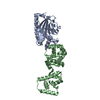
- Assembly
| Deposited unit | 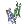
| ||||||||||||
|---|---|---|---|---|---|---|---|---|---|---|---|---|---|
| 1 | 
| ||||||||||||
| Unit cell |
|
-Components
| #1: Protein | / 102 kDa cadherin-associated protein / Alpha E-catenin / CAP102 Mass: 28830.303 Da / Num. of mol.: 2 Source method: isolated from a genetically manipulated source Source: (gene. exp.) Mus musculus (house mouse) / Gene: Ctnna1, Catna1 / Production host:  Escherichia coli BL21 (bacteria) / References: UniProt: P26231 Escherichia coli BL21 (bacteria) / References: UniProt: P26231#2: Chemical | ChemComp-BR / Bromide  Mass: 79.904 Da / Num. of mol.: 5 / Source method: obtained synthetically / Formula: Br Mass: 79.904 Da / Num. of mol.: 5 / Source method: obtained synthetically / Formula: Br#3: Chemical | Sulfate  Mass: 96.063 Da / Num. of mol.: 2 / Source method: obtained synthetically / Formula: SO4 Mass: 96.063 Da / Num. of mol.: 2 / Source method: obtained synthetically / Formula: SO4#4: Water | ChemComp-HOH / | Water Mass: 18.015 Da / Num. of mol.: 97 / Source method: isolated from a natural source / Formula: H2O Mass: 18.015 Da / Num. of mol.: 97 / Source method: isolated from a natural source / Formula: H2O |
|---|
-Experimental details
-Experiment
| Experiment | Method: X-RAY DIFFRACTION / Number of used crystals: 1 |
|---|
- Sample preparation
| Crystal | Density Matthews: 2.06 Å3/Da / Density % sol: 40.16 % |
|---|---|
| Crystal grow | Temperature: 277 K / Method: vapor diffusion, sitting drop Details: 0.2 M potassium bromide, 2.2 M ammonium sulfate, 3% w/v D-galactose |
-Data collection
| Diffraction | Mean temperature: 100 K | |||||||||
|---|---|---|---|---|---|---|---|---|---|---|
| Diffraction source | Source: SYNCHROTRON / Site: CLSI / Beamline: 08ID-1 / Wavelength: 1.0, 0.9198 | |||||||||
| Detector | Type: RAYONIX MX-300 / Detector: CCD / Date: Apr 15, 2016 | |||||||||
| Radiation | Protocol: SINGLE WAVELENGTH / Monochromatic (M) / Laue (L): M / Scattering type: x-ray | |||||||||
| Radiation wavelength |
| |||||||||
| Reflection | Resolution: 2.2→46.15 Å / Num. obs: 24938 / % possible obs: 100 % / Redundancy: 7.4 % / Biso Wilson estimate: 41.66 Å2 / CC1/2: 0.99 / Rmerge(I) obs: 0.059 / Net I/σ(I): 31.7 | |||||||||
| Reflection shell | Resolution: 2.2→2.28 Å / Redundancy: 7.4 % / Rmerge(I) obs: 0.522 / Mean I/σ(I) obs: 3.8 / CC1/2: 0.93 / % possible all: 100 |
- Processing
| Software |
| |||||||||||||||||||||||||||||||||||||||||||||||||||||||||||||||||||||||||||||||||||||||||||||||||||||||||||||||||||||||
|---|---|---|---|---|---|---|---|---|---|---|---|---|---|---|---|---|---|---|---|---|---|---|---|---|---|---|---|---|---|---|---|---|---|---|---|---|---|---|---|---|---|---|---|---|---|---|---|---|---|---|---|---|---|---|---|---|---|---|---|---|---|---|---|---|---|---|---|---|---|---|---|---|---|---|---|---|---|---|---|---|---|---|---|---|---|---|---|---|---|---|---|---|---|---|---|---|---|---|---|---|---|---|---|---|---|---|---|---|---|---|---|---|---|---|---|---|---|---|---|---|
| Refinement | Method to determine structure: SAD / Resolution: 2.2→46.15 Å / SU ML: 0.2668 / Cross valid method: FREE R-VALUE / σ(F): 0 / Phase error: 27.3052
| |||||||||||||||||||||||||||||||||||||||||||||||||||||||||||||||||||||||||||||||||||||||||||||||||||||||||||||||||||||||
| Solvent computation | Shrinkage radii: 0.9 Å / VDW probe radii: 1.11 Å | |||||||||||||||||||||||||||||||||||||||||||||||||||||||||||||||||||||||||||||||||||||||||||||||||||||||||||||||||||||||
| Displacement parameters | Biso mean: 51.32 Å2 | |||||||||||||||||||||||||||||||||||||||||||||||||||||||||||||||||||||||||||||||||||||||||||||||||||||||||||||||||||||||
| Refinement step | Cycle: LAST / Resolution: 2.2→46.15 Å
| |||||||||||||||||||||||||||||||||||||||||||||||||||||||||||||||||||||||||||||||||||||||||||||||||||||||||||||||||||||||
| Refine LS restraints |
| |||||||||||||||||||||||||||||||||||||||||||||||||||||||||||||||||||||||||||||||||||||||||||||||||||||||||||||||||||||||
| LS refinement shell |
|
