 Movie
Movie Controller


+ Open data

- Basic information
| Entry | Database: PDB / ID: 6xl3 | |||||||||
|---|---|---|---|---|---|---|---|---|---|---|
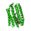
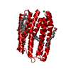
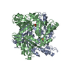
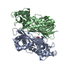
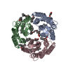
| Title | Mastigocladopsis repens rhodopsin chloride pump | |||||||||
 Components Components | Mastigocladopsis repens rhodopsin chloride pump | |||||||||
 Keywords Keywords |  MEMBRANE PROTEIN / retinal protein / proton pump / ion pump MEMBRANE PROTEIN / retinal protein / proton pump / ion pump | |||||||||
| Function / homology | TETRADECANE / DECANE / RETINAL Function and homology information Function and homology information | |||||||||
| Biological species |  Mastigocladopsis repens (bacteria) Mastigocladopsis repens (bacteria) | |||||||||
| Method | X-RAY DIFFRACTION / SYNCHROTRON / MOLECULAR REPLACEMENT / Resolution: 2.33 Å | |||||||||
 Authors Authors | Besaw, J.E. / Ernst, O.P. / Ou, W. / Morizumi, T. | |||||||||
| Funding support |  Canada, 1items Canada, 1items
| |||||||||
 Citation Citation | Journal: J.Biol.Chem. / Year: 2020 Title: The crystal structures of a chloride-pumping microbial rhodopsin and its proton-pumping mutant illuminate proton transfer determinants. Authors: Besaw, J.E. / Ou, W.L. / Morizumi, T. / Eger, B.T. / Sanchez Vasquez, J.D. / Chu, J.H.Y. / Harris, A. / Brown, L.S. / Miller, R.J.D. / Ernst, O.P. | |||||||||
| History |
|
- Structure visualization
| Structure viewer | Molecule: MolmilJmol/JSmol |
|---|
- Downloads & links
-Download
| PDBx/mmCIF format | 6xl3.cif.gz | 124.9 KB | Display | PDBx/mmCIF format |
|---|---|---|---|---|
| PDB format | pdb6xl3.ent.gz | 78.1 KB | Display | PDB format |
| PDBx/mmJSON format | 6xl3.json.gz | Tree view | PDBx/mmJSON format | |
| Others |  Other downloads Other downloads |
-Validation report
| Arichive directory | https://data.pdbj.org/pub/pdb/validation_reports/xl/6xl3ftp://data.pdbj.org/pub/pdb/validation_reports/xl/6xl3 | HTTPS FTP |
|---|
-Related structure data


-Links
 PDBj
PDBj


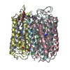
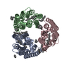
- Assembly
| Deposited unit | 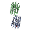
| ||||||||||||
|---|---|---|---|---|---|---|---|---|---|---|---|---|---|
| 1 | 
| ||||||||||||
| 2 | 
| ||||||||||||
| Unit cell |
|
-Components
-Protein / Sugars , 2 types, 3 molecules AB
| #1: Protein | Mass: 27131.682 Da / Num. of mol.: 2 Source method: isolated from a genetically manipulated source Source: (gene. exp.) Mastigocladopsis repens (bacteria) / Production host: Escherichia coli (E. coli)#5: Sugar | ChemComp-BOG / | Octyl glucoside Type: D-saccharide / Mass: 292.369 Da / Num. of mol.: 1 Type: D-saccharide / Mass: 292.369 Da / Num. of mol.: 1Source method: isolated from a genetically manipulated source Formula: C14H28O6 / Comment: detergent*YM |
|---|
-Non-polymers , 5 types, 53 molecules 


| #2: Chemical | Retinal Mass: 284.436 Da / Num. of mol.: 2 / Source method: obtained synthetically / Formula: C20H28O Mass: 284.436 Da / Num. of mol.: 2 / Source method: obtained synthetically / Formula: C20H28O#3: Chemical | Chloride Mass: 35.453 Da / Num. of mol.: 3 / Source method: obtained synthetically / Formula: Cl Mass: 35.453 Da / Num. of mol.: 3 / Source method: obtained synthetically / Formula: Cl#4: Chemical | Tetradecane Mass: 198.388 Da / Num. of mol.: 2 / Source method: obtained synthetically / Formula: C14H30 Mass: 198.388 Da / Num. of mol.: 2 / Source method: obtained synthetically / Formula: C14H30#6: Chemical | ChemComp-D10 / | Decane Mass: 142.282 Da / Num. of mol.: 1 / Source method: obtained synthetically / Formula: C10H22 Mass: 142.282 Da / Num. of mol.: 1 / Source method: obtained synthetically / Formula: C10H22#7: Water | ChemComp-HOH / | WaterMass: 18.015 Da / Num. of mol.: 45 / Source method: isolated from a natural source / Formula: H2O |
|---|
-Details
| Has ligand of interest | N |
|---|
-Experimental details
-Experiment
| Experiment | Method: X-RAY DIFFRACTION / Number of used crystals: 1 |
|---|
- Sample preparation
| Crystal | Density Matthews: 3.66 Å3/Da / Density % sol: 66.41 % |
|---|---|
| Crystal grow | Temperature: 307.15 K / Method: vapor diffusion, hanging drop / pH: 4 Details: 3.6 M sodium phosphate monobasic monohydrate, pH 4.0, 180 mM 1,6 hexanediol, 3.5% triethylene glycol PH range: 4.0 - 4.3 |
-Data collection
| Diffraction | Mean temperature: 100 K / Serial crystal experiment: N |
|---|---|
| Diffraction source | Source: SYNCHROTRON / Site: APS  / Beamline: 23-ID-B / Wavelength: 1.033 Å / Beamline: 23-ID-B / Wavelength: 1.033 Å |
| Detector | Type: DECTRIS EIGER X 16M / Detector: PIXEL / Date: Jul 13, 2017 |
| Radiation | Protocol: SINGLE WAVELENGTH / Monochromatic (M) / Laue (L): M / Scattering type: x-ray |
| Radiation wavelength | Wavelength: 1.033 Å / Relative weight: 1 |
| Reflection | Resolution: 2.33→27.64 Å / Num. obs: 34069 / % possible obs: 99.4 % / Redundancy: 10.3 % / Biso Wilson estimate: 35.1 Å2 / CC1/2: 0.99 / Net I/σ(I): 7.6 |
| Reflection shell | Resolution: 2.33→2.41 Å / Redundancy: 10.6 % / Mean I/σ(I) obs: 1.8 / Num. unique obs: 3344 / CC1/2: 0.36 / % possible all: 100 |
- Processing
| Software |
| |||||||||||||||||||||||||||||||||||||||||||||||||||||||||||||||||||||||||||||||||||||||||||||||||||||||||
|---|---|---|---|---|---|---|---|---|---|---|---|---|---|---|---|---|---|---|---|---|---|---|---|---|---|---|---|---|---|---|---|---|---|---|---|---|---|---|---|---|---|---|---|---|---|---|---|---|---|---|---|---|---|---|---|---|---|---|---|---|---|---|---|---|---|---|---|---|---|---|---|---|---|---|---|---|---|---|---|---|---|---|---|---|---|---|---|---|---|---|---|---|---|---|---|---|---|---|---|---|---|---|---|---|---|---|
| Refinement | Method to determine structure: MOLECULAR REPLACEMENT Starting model: IUAZ Resolution: 2.33→27.64 Å / SU ML: 0.233 / Cross valid method: FREE R-VALUE / σ(F): 1.33 / Phase error: 20.4762 Stereochemistry target values: GeoStd + Monomer Library + CDL v1.2
| |||||||||||||||||||||||||||||||||||||||||||||||||||||||||||||||||||||||||||||||||||||||||||||||||||||||||
| Solvent computation | Shrinkage radii: 0.9 Å / VDW probe radii: 1.11 Å / Solvent model: FLAT BULK SOLVENT MODEL | |||||||||||||||||||||||||||||||||||||||||||||||||||||||||||||||||||||||||||||||||||||||||||||||||||||||||
| Displacement parameters | Biso mean: 42.62 Å2 | |||||||||||||||||||||||||||||||||||||||||||||||||||||||||||||||||||||||||||||||||||||||||||||||||||||||||
| Refinement step | Cycle: LAST / Resolution: 2.33→27.64 Å
| |||||||||||||||||||||||||||||||||||||||||||||||||||||||||||||||||||||||||||||||||||||||||||||||||||||||||
| Refine LS restraints |
| |||||||||||||||||||||||||||||||||||||||||||||||||||||||||||||||||||||||||||||||||||||||||||||||||||||||||
| LS refinement shell |
|
