 Movie
Movie Controller


+ Open data

- Basic information
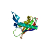
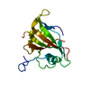
| Entry | Database: PDB / ID: 3dj1 | ||||||
|---|---|---|---|---|---|---|---|
| Title | crystal structure of TIP-1 wild type | ||||||
 Components Components | Tax1-binding protein 3 | ||||||
 Keywords Keywords |  SIGNALING PROTEIN / Tax-interacting protein-1 / PDZ domain / TIP-1 / Cytoplasm / Nucleus / Wnt signaling pathway SIGNALING PROTEIN / Tax-interacting protein-1 / PDZ domain / TIP-1 / Cytoplasm / Nucleus / Wnt signaling pathway | ||||||
| Function / homology |  Function and homology information Function and homology informationRHO GTPases Activate Rhotekin and Rhophilins / negative regulation of protein localization to cell surface / negative regulation of Wnt signaling pathway / Rho protein signal transduction / fibrillar center / beta-catenin binding / Wnt signaling pathway / actin cytoskeleton / negative regulation of cell population proliferation / intracellular membrane-bounded organelle ...RHO GTPases Activate Rhotekin and Rhophilins / negative regulation of protein localization to cell surface / negative regulation of Wnt signaling pathway / Rho protein signal transduction / fibrillar center / beta-catenin binding / Wnt signaling pathway / actin cytoskeleton / negative regulation of cell population proliferation / intracellular membrane-bounded organelle / nucleus / plasma membrane / cytoplasmSimilarity search - Function | ||||||
| Biological species |  Mus musculus (house mouse) Mus musculus (house mouse) | ||||||
| Method | X-RAY DIFFRACTION / MOLECULAR REPLACEMENT / Resolution: 1.8 Å | ||||||
 Authors Authors | Shen, Y. | ||||||
 Citation Citation | Journal: J.Mol.Biol. / Year: 2008 Title: Structural Basis of beta-Catenin Recognition by Tax-interacting Protein-1 Authors: Zhang, J. / Yan, X. / Shi, C. / Yang, X. / Guo, Y. / Tian, C. / Long, J. / Shen, Y. | ||||||
| History |
|
- Structure visualization
| Structure viewer | Molecule: MolmilJmol/JSmol |
|---|
- Downloads & links
-Download
| PDBx/mmCIF format | 3dj1.cif.gz | 59.2 KB | Display | PDBx/mmCIF format |
|---|---|---|---|---|
| PDB format | pdb3dj1.ent.gz | 42.8 KB | Display | PDB format |
| PDBx/mmJSON format | 3dj1.json.gz | Tree view | PDBx/mmJSON format | |
| Others |  Other downloads Other downloads |
-Validation report
| Arichive directory | https://data.pdbj.org/pub/pdb/validation_reports/dj/3dj1ftp://data.pdbj.org/pub/pdb/validation_reports/dj/3dj1 | HTTPS FTP |
|---|
-Related structure data
| Related structure data |  3diwSC  3dj3C S: Starting model for refinement C: citing same article ( |
|---|---|
| Similar structure data |


-Links
 PDBj
PDBj- Assembly
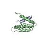
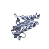
| Deposited unit | 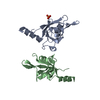
| ||||||||||||
|---|---|---|---|---|---|---|---|---|---|---|---|---|---|
| 1 | 
| ||||||||||||
| 2 | 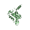
| ||||||||||||
| 3 | 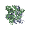
| ||||||||||||
| Unit cell |
| ||||||||||||
| Components on special symmetry positions |
|
-Components
| #1: Protein | Mass: 13739.631 Da / Num. of mol.: 2 Source method: isolated from a genetically manipulated source Source: (gene. exp.) Mus musculus (house mouse) / Production host:  Escherichia coli (E. coli) / References: UniProt: Q9DBG9 Escherichia coli (E. coli) / References: UniProt: Q9DBG9#2: Chemical | ChemComp-SO4 / | Sulfate  Mass: 96.063 Da / Num. of mol.: 1 / Source method: obtained synthetically / Formula: SO4 Mass: 96.063 Da / Num. of mol.: 1 / Source method: obtained synthetically / Formula: SO4#3: Water | ChemComp-HOH / | Water Mass: 18.015 Da / Num. of mol.: 136 / Source method: isolated from a natural source / Formula: H2O Mass: 18.015 Da / Num. of mol.: 136 / Source method: isolated from a natural source / Formula: H2O |
|---|
-Experimental details
-Experiment
| Experiment | Method: X-RAY DIFFRACTION / Number of used crystals: 1 |
|---|
- Sample preparation
| Crystal | Density Matthews: 2.12 Å3/Da / Density % sol: 41.9 % |
|---|---|
| Crystal grow | Temperature: 293 K / Method: vapor diffusion, hanging drop / pH: 9 Details: 1.6M Ammonium sulfate, 0.1M bicine buffer, pH 9.0, VAPOR DIFFUSION, HANGING DROP, temperature 293K |
-Data collection
| Diffraction | Mean temperature: 100 K |
|---|---|
| Diffraction source | Source: ROTATING ANODE / Type: RIGAKU MICROMAX-007 / Wavelength: 1.54 Å |
| Detector | Type: RIGAKU RAXIS IV++ / Detector: IMAGE PLATE / Date: Nov 6, 2007 |
| Radiation | Protocol: SINGLE WAVELENGTH / Monochromatic (M) / Laue (L): M / Scattering type: x-ray |
| Radiation wavelength | Wavelength: 1.54 Å / Relative weight: 1 |
| Reflection | Resolution: 1.8→50 Å / Num. all: 21968 / Num. obs: 21858 / % possible obs: 99.5 % / Observed criterion σ(F): 2 / Observed criterion σ(I): 2 / Redundancy: 5.4 % / Biso Wilson estimate: 23.2 Å2 / Rsym value: 0.025 / Net I/σ(I): 63.3 |
| Reflection shell | Resolution: 1.77→1.83 Å / Redundancy: 4.6 % / Rmerge(I) obs: 0.124 / Mean I/σ(I) obs: 13.2 / Num. unique all: 2104 / % possible all: 95.7 |
- Processing
| Software |
| ||||||||||||||||||||||||||||||||||||
|---|---|---|---|---|---|---|---|---|---|---|---|---|---|---|---|---|---|---|---|---|---|---|---|---|---|---|---|---|---|---|---|---|---|---|---|---|---|
| Refinement | Method to determine structure: MOLECULAR REPLACEMENT Starting model: PDB ENTRY 3DIW Resolution: 1.8→27.44 Å / Rfactor Rfree error: 0.005 / Data cutoff high absF: 1241665.59 / Data cutoff low absF: 0 / Isotropic thermal model: RESTRAINED / Cross valid method: THROUGHOUT / σ(F): 0 / Stereochemistry target values: Engh & Huber / Details: BULK SOLVENT MODEL USED
| ||||||||||||||||||||||||||||||||||||
| Solvent computation | Solvent model: FLAT MODEL / Bsol: 38.8231 Å2 / ksol: 0.4 e/Å3 | ||||||||||||||||||||||||||||||||||||
| Displacement parameters | Biso mean: 27.1 Å2
| ||||||||||||||||||||||||||||||||||||
| Refine analyze |
| ||||||||||||||||||||||||||||||||||||
| Refinement step | Cycle: LAST / Resolution: 1.8→27.44 Å
| ||||||||||||||||||||||||||||||||||||
| Refine LS restraints |
| ||||||||||||||||||||||||||||||||||||
| LS refinement shell | Resolution: 1.8→1.91 Å / Rfactor Rfree error: 0.018 / Total num. of bins used: 6
| ||||||||||||||||||||||||||||||||||||
| Xplor file |
|
