 Movie
Movie Controller


+ Open data

- Basic information
| Entry | Database: PDB / ID: 5hd2 | ||||||
|---|---|---|---|---|---|---|---|
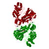
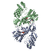
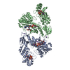
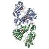
| Title | The crystal structure of SeMet-Cry51Aa2-L11M | ||||||
 Components Components | Parasporal crystal protein | ||||||
 Keywords Keywords |  TOXIN / Cry51Aa2 / Bt / Cry TOXIN / Cry51Aa2 / Bt / Cry | ||||||
| Function / homology | Aerolysin-like toxin / Clostridium epsilon toxin ETX/Bacillus mosquitocidal toxin MTX2 / Parasporal crystal protein Function and homology information Function and homology information | ||||||
| Biological species |  Bacillus thuringiensis (bacteria) Bacillus thuringiensis (bacteria) | ||||||
| Method | X-RAY DIFFRACTION / SYNCHROTRON / MAD / Resolution: 2.276 Å | ||||||
 Authors Authors | Rydel, T.J. / Sturman, E.J. / Moshiri, F. | ||||||
 Citation Citation | Journal: Nat Commun / Year: 2016 Title: A transgenic approach for controlling Lygus in cotton. Authors: Gowda, A. / Rydel, T.J. / Wollacott, A.M. / Brown, R.S. / Akbar, W. / Clark, T.L. / Flasinski, S. / Nageotte, J.R. / Read, A.C. / Shi, X. / Werner, B.J. / Pleau, M.J. / Baum, J.A. | ||||||
| History |
|
- Structure visualization
| Structure viewer | Molecule: MolmilJmol/JSmol |
|---|
- Downloads & links
-Download
| PDBx/mmCIF format | 5hd2.cif.gz | 70.1 KB | Display | PDBx/mmCIF format |
|---|---|---|---|---|
| PDB format | pdb5hd2.ent.gz | 55.3 KB | Display | PDB format |
| PDBx/mmJSON format | 5hd2.json.gz | Tree view | PDBx/mmJSON format | |
| Others |  Other downloads Other downloads |
-Validation report
| Arichive directory | https://data.pdbj.org/pub/pdb/validation_reports/hd/5hd2ftp://data.pdbj.org/pub/pdb/validation_reports/hd/5hd2 | HTTPS FTP |
|---|
-Related structure data
| Similar structure data |
|---|


-Links
 PDBj
PDBj- Assembly
| Deposited unit | 
| ||||||||
|---|---|---|---|---|---|---|---|---|---|
| 1 | 
| ||||||||
| Unit cell |
|
-Components
| #1: Protein | Mass: 34356.023 Da / Num. of mol.: 1 / Mutation: L11M Source method: isolated from a genetically manipulated source Source: (gene. exp.) Bacillus thuringiensis (bacteria) / Plasmid: pET28a + pRARE2 / Production host: Escherichia coli (E. coli) / Strain (production host): B834(DE3) / References: UniProt: E9KBU4 |
|---|---|
| #2: Water | ChemComp-HOH / Water Mass: 18.015 Da / Num. of mol.: 47 / Source method: isolated from a natural source / Formula: H2O Mass: 18.015 Da / Num. of mol.: 47 / Source method: isolated from a natural source / Formula: H2O |
-Experimental details
-Experiment
| Experiment | Method: X-RAY DIFFRACTION |
|---|
- Sample preparation
| Crystal | Density Matthews: 2.37 Å3/Da / Density % sol: 48 % / Description: Bipyramidal. |
|---|---|
| Crystal grow | Temperature: 293 K / Method: vapor diffusion, sitting drop / pH: 7.5 Details: Starting protein solution was 5.5 mg/ml protein in 25 mM sodium carbonate buffer-pH 10.5. The reservoir solution was 500 ul of 2.0 M sodium chloride and 50 mM HEPES-pH 7.5 buffer. ...Details: Starting protein solution was 5.5 mg/ml protein in 25 mM sodium carbonate buffer-pH 10.5. The reservoir solution was 500 ul of 2.0 M sodium chloride and 50 mM HEPES-pH 7.5 buffer. Bipyramidal crystals resulted from 2 ul drops, using 0.7 ul protein solution and 1.3 ul well solution. PH range: ~7.5 / Temp details: room temperature |
-Data collection
| Diffraction | Mean temperature: 100 K Ambient temp details: The structure was initially solved using four wavelengths of SeMet MAD data to 2.8 A resolution. The structure was extended to 2.27 A using data collected at 2.27 A resolution ...Ambient temp details: The structure was initially solved using four wavelengths of SeMet MAD data to 2.8 A resolution. The structure was extended to 2.27 A using data collected at 2.27 A resolution on the SER-CAT 22-ID line. |
|---|---|
| Diffraction source | Source: SYNCHROTRON / Site: APS  / Beamline: 21-ID-D / Wavelength: 1 Å / Beamline: 21-ID-D / Wavelength: 1 Å |
| Detector | Type: MAR scanner 300 mm plate / Detector: IMAGE PLATE / Date: Dec 12, 2007 Details: 115 data frames were collected using 3 second exposures, with an oscillation angle of 1 degree, and a crystal-to-detector distance of 200 mm. |
| Radiation | Protocol: SINGLE WAVELENGTH / Monochromatic (M) / Laue (L): M / Scattering type: x-ray |
| Radiation wavelength | Wavelength: 1 Å / Relative weight: 1 |
| Reflection | Resolution: 2.27→53.7 Å / Num. all: 15236 / Num. obs: 15236 / % possible obs: 95.9 % / Redundancy: 4.7 % / Rmerge(I) obs: 0.094 / Net I/σ(I): 17.4 |
| Reflection shell | Resolution: 2.27→2.35 Å / Redundancy: 4.9 % / Rmerge(I) obs: 0.381 / Mean I/σ(I) obs: 1.75 / % possible all: 96.4 |
- Processing
| Software |
| ||||||||||||||||||||||||||||||||||||||||||||||||||||||||||||||||||||||||||||||||||||||||||||||||||||||||||||||||||||||||||||||||||||||||||||||||||||||||||||||||||||||||||||||||||||||
|---|---|---|---|---|---|---|---|---|---|---|---|---|---|---|---|---|---|---|---|---|---|---|---|---|---|---|---|---|---|---|---|---|---|---|---|---|---|---|---|---|---|---|---|---|---|---|---|---|---|---|---|---|---|---|---|---|---|---|---|---|---|---|---|---|---|---|---|---|---|---|---|---|---|---|---|---|---|---|---|---|---|---|---|---|---|---|---|---|---|---|---|---|---|---|---|---|---|---|---|---|---|---|---|---|---|---|---|---|---|---|---|---|---|---|---|---|---|---|---|---|---|---|---|---|---|---|---|---|---|---|---|---|---|---|---|---|---|---|---|---|---|---|---|---|---|---|---|---|---|---|---|---|---|---|---|---|---|---|---|---|---|---|---|---|---|---|---|---|---|---|---|---|---|---|---|---|---|---|---|---|---|---|---|
| Refinement | Method to determine structure: MAD / Resolution: 2.276→53.7 Å / Cor.coef. Fo:Fc: 0.93 / Cor.coef. Fo:Fc free: 0.899 / SU B: 8.299 / SU ML: 0.208 / Cross valid method: THROUGHOUT / ESU R: 0.394 / ESU R Free: 0.283 / Stereochemistry target values: MAXIMUM LIKELIHOOD / Details: HYDROGENS HAVE BEEN ADDED IN THE RIDING POSITIONS
| ||||||||||||||||||||||||||||||||||||||||||||||||||||||||||||||||||||||||||||||||||||||||||||||||||||||||||||||||||||||||||||||||||||||||||||||||||||||||||||||||||||||||||||||||||||||
| Solvent computation | Ion probe radii: 0.8 Å / Shrinkage radii: 0.8 Å / VDW probe radii: 1.4 Å / Solvent model: MASK | ||||||||||||||||||||||||||||||||||||||||||||||||||||||||||||||||||||||||||||||||||||||||||||||||||||||||||||||||||||||||||||||||||||||||||||||||||||||||||||||||||||||||||||||||||||||
| Displacement parameters | Biso mean: 41.305 Å2
| ||||||||||||||||||||||||||||||||||||||||||||||||||||||||||||||||||||||||||||||||||||||||||||||||||||||||||||||||||||||||||||||||||||||||||||||||||||||||||||||||||||||||||||||||||||||
| Refinement step | Cycle: LAST / Resolution: 2.276→53.7 Å
| ||||||||||||||||||||||||||||||||||||||||||||||||||||||||||||||||||||||||||||||||||||||||||||||||||||||||||||||||||||||||||||||||||||||||||||||||||||||||||||||||||||||||||||||||||||||
| Refine LS restraints |
|