 Movie
Movie Controller


+ Open data

- Basic information
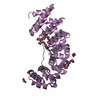
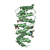
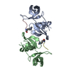
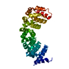
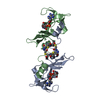
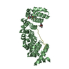
| Entry | Database: PDB / ID: 1nf1 | ||||||
|---|---|---|---|---|---|---|---|
| Title | THE GAP RELATED DOMAIN OF NEUROFIBROMIN | ||||||
 Components Components | PROTEIN (NEUROFIBROMIN) | ||||||
 Keywords Keywords |  SIGNALING PROTEIN / NEUROFIBROMIN / TYPE I NEUROFIBROMATOSIS / NF1 / RAS / GAP / SIGNAL TRANSDUCTION / CANCER / GROWTH REGULATION / GTP HYDROLYSIS / PATIENT MUTATION / ARGININE FINGER SIGNALING PROTEIN / NEUROFIBROMIN / TYPE I NEUROFIBROMATOSIS / NF1 / RAS / GAP / SIGNAL TRANSDUCTION / CANCER / GROWTH REGULATION / GTP HYDROLYSIS / PATIENT MUTATION / ARGININE FINGER | ||||||
| Function / homology |  Function and homology information Function and homology informationpositive regulation of mast cell apoptotic process / negative regulation of Rac protein signal transduction / regulation of glial cell differentiation / gamma-aminobutyric acid secretion, neurotransmission / observational learning / Schwann cell migration / negative regulation of Schwann cell migration / vascular associated smooth muscle cell migration / amygdala development / mast cell apoptotic process ...positive regulation of mast cell apoptotic process / negative regulation of Rac protein signal transduction / regulation of glial cell differentiation / gamma-aminobutyric acid secretion, neurotransmission / observational learning / Schwann cell migration / negative regulation of Schwann cell migration / vascular associated smooth muscle cell migration / amygdala development / mast cell apoptotic process / negative regulation of mast cell proliferation / Schwann cell proliferation / vascular associated smooth muscle cell proliferation / mast cell proliferation / glutamate secretion, neurotransmission / negative regulation of Schwann cell proliferation / negative regulation of leukocyte migration / negative regulation of vascular associated smooth muscle cell migration / positive regulation of adenylate cyclase activity / regulation of cell-matrix adhesion / forebrain morphogenesis / negative regulation of neurotransmitter secretion / hair follicle maturation / cell communication / regulation of blood vessel endothelial cell migration / camera-type eye morphogenesis / smooth muscle tissue development / negative regulation of oligodendrocyte differentiation / sympathetic nervous system development / myelination in peripheral nervous system / myeloid leukocyte migration / phosphatidylcholine binding / peripheral nervous system development / phosphatidylethanolamine binding / metanephros development / positive regulation of extrinsic apoptotic signaling pathway in absence of ligand / negative regulation of Ras protein signal transduction / collagen fibril organization / regulation of bone resorption / regulation of long-term synaptic potentiation / neural tube development / endothelial cell proliferation / forebrain astrocyte development / pigmentation / artery morphogenesis / regulation of postsynapse organization / regulation of synaptic transmission, GABAergic / negative regulation of neuroblast proliferation / negative regulation of MAPK cascade / adrenal gland development / negative regulation of protein import into nucleus / negative regulation of cell-matrix adhesion / spinal cord development / regulation of GTPase activity / Rac protein signal transduction / oligodendrocyte differentiation / negative regulation of osteoclast differentiation / negative regulation of endothelial cell proliferation / RAS signaling downstream of NF1 loss-of-function variants / negative regulation of astrocyte differentiation / extrinsic apoptotic signaling pathway via death domain receptors / neuroblast proliferation / regulation of angiogenesis / Schwann cell development / negative regulation of stem cell proliferation / negative regulation of fibroblast proliferation / skeletal muscle tissue development / extrinsic apoptotic signaling pathway in absence of ligand / positive regulation of vascular associated smooth muscle cell proliferation / positive regulation of endothelial cell proliferation / extracellular matrix organization / negative regulation of angiogenesis / GTPase activator activity / negative regulation of cell migration / osteoclast differentiation / regulation of ERK1 and ERK2 cascade / phosphatidylinositol 3-kinase/protein kinase B signal transduction / liver development / negative regulation of MAP kinase activity / stem cell proliferation / long-term synaptic potentiation / regulation of long-term neuronal synaptic plasticity / negative regulation of protein kinase activity / wound healing / brain development / visual learning / cerebral cortex development / cognition / positive regulation of GTPase activity / osteoblast differentiation / Regulation of RAS by GAPs / protein import into nucleus / MAPK cascade / positive regulation of neuron apoptotic process / presynapse / heart development / cellular response to heat / fibroblast proliferation / actin cytoskeleton organization / regulation of gene expressionSimilarity search - Function | ||||||
| Biological species |  Homo sapiens (human) Homo sapiens (human) | ||||||
| Method | X-RAY DIFFRACTION / SYNCHROTRON / MIR / Resolution: 2.5 Å | ||||||
 Authors Authors | Scheffzek, K. / Ahmadian, M.R. / Wiesmueller, L. / Kabsch, W. / Stege, P. / Schmitz, F. / Wittinghofer, A. | ||||||
 Citation Citation | Journal: EMBO J. / Year: 1998 Title: Structural analysis of the GAP-related domain from neurofibromin and its implications. Authors: Scheffzek, K. / Ahmadian, M.R. / Wiesmuller, L. / Kabsch, W. / Stege, P. / Schmitz, F. / Wittinghofer, A. #1: Journal: Science / Year: 1997Title: The Ras-Rasgap Complex: Structural Basis for Gtpase Activation and its Loss in Oncogenic Ras Mutants Authors: Scheffzek, K. / Ahmadian, M.R. / Kabsch, W. / Wiesmueller, L. / Lautwein, A. / Schmitz, F. / Wittinghofer, A. #2: Journal: Nat.Struct.Biol. / Year: 1997Title: Confirmation of the Arginine-Finger Hypothesis for the Gap-Stimulated GTP- Hydrolysis Reaction of Ras Authors: Ahmadian, M.R. / Stege, P. / Scheffzek, K. / Wittinghofer, A. #3: Journal: J.Biol.Chem. / Year: 1996Title: Structural Differences in the Minimal Catalytic Domains of the Gtpasse- Activating Proteins P120Gap and Neurofibromin Authors: Ahmadian, M.R. / Wiesmueller, L. / Lautwein, A. / Bischoff, F.R. / Wittinghofer, A. #4: Journal: Nature / Year: 1996Title: 3-Dimensional Structure of the Gtpase Activating Domain of Human P120Gap and Implications for the Interaction with Ras Authors: Scheffzek, K. / Lautwein, A. / Kabsch, W. / Ahmadian, M.R. / Wittinghofer, A. #5: Journal: Science / Year: 1996Title: Formation of a Transition-State Analog of the Ras Gtpase Reaction by Ras:Gdp, Tetrafluoroaluminate and Gtpase-Activating Proteins Authors: Mittal, R. / Ahmadian, M.R. / Goody, R.S. / Wittinghofer, A. #6: Journal: Neuron / Year: 1993Title: The Neurofibromatosis Type I Gene and its Protein Product Authors: Gutmann, D.H. / Collins, F.S. #7: Journal: Cell(Cambridge,Mass.) / Year: 1990Title: The NF1 Locus Encodes a Protein Functionally Related to Mammalian Gap and Yeast Ira Proteins Authors: Ballester, R. / Marchuk, D. / Boguski, M. / Saulino, A. / Letcher, R. / Wigler, M. / Collins, F. #8: Journal: Cell(Cambridge,Mass.) / Year: 1990Title: The Gap-Related Domain of the Neurofibromatosis Type I Gene Product Interacts with Ras P21 Authors: Martin, G.A. / Viskochil, D. / Bollag, G. / Mccabe, P.C. / Crosier, W.J. / Hausbruck, H. / Conroy, L. / Clark, R. / O'Connell, P. / Cawthon, R.M. / Innis, M.A. / Mccormick, F. #9: Journal: Cell(Cambridge,Mass.) / Year: 1990Title: The Catalytic Domain of the Neurofibromatosis Type I Gene Product Stimulates Ras Gtpase and Complements Ira Mutants of S. Cerevisiae Authors: Xu, G. / Lin, B. / Tanaka, K. / Dunn, D. / Wod, D. / Gesteland, R. / White, R. / Weiss, R. / Tamanoi, F. #10: Journal: Embo J. / Year: 1990Title: Refined Crystal Structure of the Triphosphate Conformation of H-Ras P21 at 1.35 A Resolution: Implications for the Mechanism of GTP Hydrolysis Authors: Pai, E.F. / Krengel, U. / Petsko, G.A. / Goody, R.S. / Kabsch, W. / Wittinghofer, A. | ||||||
| History |
|
- Structure visualization
| Structure viewer | Molecule: MolmilJmol/JSmol |
|---|
- Downloads & links
-Download
| PDBx/mmCIF format | 1nf1.cif.gz | 64.6 KB | Display | PDBx/mmCIF format |
|---|---|---|---|---|
| PDB format | pdb1nf1.ent.gz | 46.8 KB | Display | PDB format |
| PDBx/mmJSON format | 1nf1.json.gz | Tree view | PDBx/mmJSON format | |
| Others |  Other downloads Other downloads |
-Validation report
| Arichive directory | https://data.pdbj.org/pub/pdb/validation_reports/nf/1nf1ftp://data.pdbj.org/pub/pdb/validation_reports/nf/1nf1 | HTTPS FTP |
|---|
-Related structure data
| Similar structure data |
|---|


-Links
 PDBj
PDBj


- Assembly
| Deposited unit | 
| ||||||||
|---|---|---|---|---|---|---|---|---|---|
| 1 |
| ||||||||
| Unit cell |
|
-Components
| #1: Protein | Mass: 37982.672 Da / Num. of mol.: 1 / Fragment: GAP RELATED DOMAIN Source method: isolated from a genetically manipulated source Details: SEE REF.5 FOR DETAILS / Source: (gene. exp.) Homo sapiens (human) / Description: S. REF. 4 / Cellular location: CYTOSOL / Plasmid: PETNF1-333 / Production host:  Escherichia coli (E. coli) / Strain (production host): DG103 / References: UniProt: P21359 Escherichia coli (E. coli) / Strain (production host): DG103 / References: UniProt: P21359 |
|---|
-Experimental details
-Experiment
| Experiment | Method: X-RAY DIFFRACTION / Number of used crystals: 6 |
|---|
- Sample preparation
| Crystal | Density Matthews: 2.24 Å3/Da / Density % sol: 44.98 % | ||||||||||||||||||||||||||||||||||||||||||||||||
|---|---|---|---|---|---|---|---|---|---|---|---|---|---|---|---|---|---|---|---|---|---|---|---|---|---|---|---|---|---|---|---|---|---|---|---|---|---|---|---|---|---|---|---|---|---|---|---|---|---|
| Crystal grow | pH: 8 / Details: S. REF. DESCRIBING THE STRUCTURE, pH 8 | ||||||||||||||||||||||||||||||||||||||||||||||||
| Crystal grow | *PLUS pH: 7.5 / Method: vapor diffusion, hanging drop | ||||||||||||||||||||||||||||||||||||||||||||||||
| Components of the solutions | *PLUS
|
-Data collection
| Diffraction | Mean temperature: 277 K |
|---|---|
| Diffraction source | Source: SYNCHROTRON / Site: EMBL/DESY, HAMBURG  / Beamline: X11 / Wavelength: 0.91 / Beamline: X11 / Wavelength: 0.91 |
| Detector | Type: MARRESEARCH / Detector: IMAGE PLATE / Date: Feb 26, 1997 |
| Radiation | Protocol: SINGLE WAVELENGTH / Monochromatic (M) / Laue (L): M / Scattering type: x-ray |
| Radiation wavelength | Wavelength: 0.91 Å / Relative weight: 1 |
| Reflection | Resolution: 2.5→100 Å / Num. obs: 50172 / % possible obs: 99 % / Redundancy: 4.3 % / Biso Wilson estimate: 52.4 Å2 / Rmerge(I) obs: 0.07 / Net I/σ(I): 11.6 |
| Reflection shell | Resolution: 2.5→2.6 Å / Redundancy: 3.1 % / Rmerge(I) obs: 0.3 / Mean I/σ(I) obs: 3.6 / % possible all: 99.8 |
| Reflection | *PLUS Num. obs: 11665 / % possible obs: 99.8 % / Num. measured all: 50172 / Rmerge(I) obs: 0.073 |
- Processing
| Software |
| ||||||||||||||||||||||||||||||||||||||||||||||||||||||||||||||||||||||||||||||||
|---|---|---|---|---|---|---|---|---|---|---|---|---|---|---|---|---|---|---|---|---|---|---|---|---|---|---|---|---|---|---|---|---|---|---|---|---|---|---|---|---|---|---|---|---|---|---|---|---|---|---|---|---|---|---|---|---|---|---|---|---|---|---|---|---|---|---|---|---|---|---|---|---|---|---|---|---|---|---|---|---|---|
| Refinement | Method to determine structure: MIR / Resolution: 2.5→30 Å / Rfactor Rfree error: 0.011 / Data cutoff high absF: 100000 / Data cutoff low absF: 0.001 / Isotropic thermal model: RESTRAINED / Cross valid method: THROUGHOUT / σ(F): 2 / Details: BULK SOLVENT MODEL USED IN FINAL STAGES
| ||||||||||||||||||||||||||||||||||||||||||||||||||||||||||||||||||||||||||||||||
| Displacement parameters | Biso mean: 47.6 Å2 | ||||||||||||||||||||||||||||||||||||||||||||||||||||||||||||||||||||||||||||||||
| Refine analyze |
| ||||||||||||||||||||||||||||||||||||||||||||||||||||||||||||||||||||||||||||||||
| Refinement step | Cycle: LAST / Resolution: 2.5→30 Å
| ||||||||||||||||||||||||||||||||||||||||||||||||||||||||||||||||||||||||||||||||
| Refine LS restraints |
| ||||||||||||||||||||||||||||||||||||||||||||||||||||||||||||||||||||||||||||||||
| LS refinement shell | Resolution: 2.5→2.61 Å / Rfactor Rfree error: 0.036 / Total num. of bins used: 8
| ||||||||||||||||||||||||||||||||||||||||||||||||||||||||||||||||||||||||||||||||
| Software | *PLUS Name: X-PLOR / Version: 3.8 / Classification: refinement | ||||||||||||||||||||||||||||||||||||||||||||||||||||||||||||||||||||||||||||||||
| Refinement | *PLUS Highest resolution: 2.5 Å / Lowest resolution: 30 Å / σ(F): 2 / % reflection Rfree: 10 % | ||||||||||||||||||||||||||||||||||||||||||||||||||||||||||||||||||||||||||||||||
| Solvent computation | *PLUS | ||||||||||||||||||||||||||||||||||||||||||||||||||||||||||||||||||||||||||||||||
| Displacement parameters | *PLUS | ||||||||||||||||||||||||||||||||||||||||||||||||||||||||||||||||||||||||||||||||
| Refine LS restraints | *PLUS
| ||||||||||||||||||||||||||||||||||||||||||||||||||||||||||||||||||||||||||||||||
| LS refinement shell | *PLUS Highest resolution: 2.5 Å / Rfactor Rfree: 0.436 / % reflection Rfree: 11.9 % / Rfactor Rwork: 0.4 |
