 Movie
Movie Controller

[English] 日本語
 Yorodumi
- PDB-4cnc: Crystal structure of human 5T4 (Wnt-activated inhibitory factor 1... -
+ Open data

- Basic information
| Entry | Database: PDB / ID: 4cnc | |||||||||
|---|---|---|---|---|---|---|---|---|---|---|
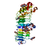
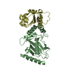
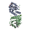
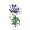
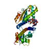
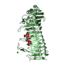
| Title | Crystal structure of human 5T4 (Wnt-activated inhibitory factor 1, Trophoblast glycoprotein) | |||||||||
 Components Components | TROPHOBLAST GLYCOPROTEIN | |||||||||
 Keywords Keywords |  CELL ADHESION / WAIF1 / TPBG / TROVAX / CANCER / SIGNALING / LEUCINE-RICH REPEATS / WNT/BETA-CATENIN SIGNALING PATHWAY CELL ADHESION / WAIF1 / TPBG / TROVAX / CANCER / SIGNALING / LEUCINE-RICH REPEATS / WNT/BETA-CATENIN SIGNALING PATHWAY | |||||||||
| Function / homology |  Function and homology information Function and homology informationmesenchymal cell migration / dendrite arborization / olfactory learning / positive regulation of chemotaxis / synaptic transmission, GABAergic / positive regulation of synapse assembly / axon terminus / cell chemotaxis / protein localization to plasma membrane / negative regulation of canonical Wnt signaling pathway ...mesenchymal cell migration / dendrite arborization / olfactory learning / positive regulation of chemotaxis / synaptic transmission, GABAergic / positive regulation of synapse assembly / axon terminus / cell chemotaxis / protein localization to plasma membrane / negative regulation of canonical Wnt signaling pathway / positive regulation of phosphatidylinositol 3-kinase/protein kinase B signal transduction / positive regulation of ERK1 and ERK2 cascade / cell adhesion / negative regulation of cell population proliferation / dendrite / cell surface / endoplasmic reticulum / plasma membraneSimilarity search - Function | |||||||||
| Biological species |  HOMO SAPIENS (human) HOMO SAPIENS (human) | |||||||||
| Method | X-RAY DIFFRACTION / SYNCHROTRON / MOLECULAR REPLACEMENT / Resolution: 1.77 Å | |||||||||
 Authors Authors | Zhao, Y. / Malinauskas, T. / Harlos, K. / Jones, E.Y. | |||||||||
 Citation Citation | Journal: Structure / Year: 2014 Title: Structural Insights Into the Inhibition of Wnt Signaling by Cancer Antigen 5T4/Wnt-Activated Inhibitory Factor 1. Authors: Zhao, Y. / Malinauskas, T. / Harlos, K. / Jones, E.Y. | |||||||||
| History |
|
- Structure visualization
| Structure viewer | Molecule: MolmilJmol/JSmol |
|---|
- Downloads & links
-Download
| PDBx/mmCIF format | 4cnc.cif.gz | 232.7 KB | Display | PDBx/mmCIF format |
|---|---|---|---|---|
| PDB format | pdb4cnc.ent.gz | 188 KB | Display | PDB format |
| PDBx/mmJSON format | 4cnc.json.gz | Tree view | PDBx/mmJSON format | |
| Others |  Other downloads Other downloads |
-Validation report
| Arichive directory | https://data.pdbj.org/pub/pdb/validation_reports/cn/4cncftp://data.pdbj.org/pub/pdb/validation_reports/cn/4cnc | HTTPS FTP |
|---|
-Related structure data


-Links
 PDBj
PDBj


- Assembly
| Deposited unit | 
| ||||||||
|---|---|---|---|---|---|---|---|---|---|
| 1 | 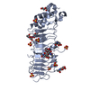
| ||||||||
| 2 | 
| ||||||||
| Unit cell |
| ||||||||
| Noncrystallographic symmetry (NCS) | NCS oper: (Code: given Matrix: (-0.9999, -0.0099, -0.0092), Vector : |
-Components
-Protein , 1 types, 2 molecules AB
| #1: Protein | Mass: 32900.395 Da / Num. of mol.: 2 / Fragment: EXTRACELLULAR DOMAIN, RESIDUES 60-345 Source method: isolated from a genetically manipulated source Source: (gene. exp.) HOMO SAPIENS (human) / Tissue: EMBRYONIC KIDNEY / Organ: KIDNEY / Plasmid: PURD-5T4/WAIF1ECTO / Cell line (production host): HEK293S GNTI- / Production host: HOMO SAPIENS (human) / References: UniProt: Q13641 |
|---|
-Sugars , 2 types, 8 molecules 
| #2: Polysaccharide | / Mass: 424.401 Da / Num. of mol.: 2 Source method: isolated from a genetically manipulated source #3: Sugar | ChemComp-NAG / N-Acetylglucosamine Type: D-saccharide, beta linking / Mass: 221.208 Da / Num. of mol.: 6 Type: D-saccharide, beta linking / Mass: 221.208 Da / Num. of mol.: 6Source method: isolated from a genetically manipulated source Formula: C8H15NO6 |
|---|
-Non-polymers , 5 types, 201 molecules 


| #4: Chemical | ChemComp-PEG / Diethylene glycol Mass: 106.120 Da / Num. of mol.: 4 / Source method: obtained synthetically / Formula: C4H10O3 Mass: 106.120 Da / Num. of mol.: 4 / Source method: obtained synthetically / Formula: C4H10O3#5: Chemical | Glycerol Mass: 92.094 Da / Num. of mol.: 3 / Source method: obtained synthetically / Formula: C3H8O3 Mass: 92.094 Da / Num. of mol.: 3 / Source method: obtained synthetically / Formula: C3H8O3#6: Chemical | ChemComp-SO4 / Sulfate Mass: 96.063 Da / Num. of mol.: 14 / Source method: obtained synthetically / Formula: SO4 Mass: 96.063 Da / Num. of mol.: 14 / Source method: obtained synthetically / Formula: SO4#7: Chemical | ChemComp-NA / |  Mass: 22.990 Da / Num. of mol.: 1 / Source method: obtained synthetically / Formula: Na Mass: 22.990 Da / Num. of mol.: 1 / Source method: obtained synthetically / Formula: Na#8: Water | ChemComp-HOH / | WaterMass: 18.015 Da / Num. of mol.: 179 / Source method: isolated from a natural source / Formula: H2O |
|---|
-Experimental details
-Experiment
| Experiment | Method: X-RAY DIFFRACTION / Number of used crystals: 1 |
|---|
- Sample preparation
| Crystal | Density Matthews: 1.8 Å3/Da / Density % sol: 33 % / Description: NONE |
|---|---|
| Crystal grow | Method: vapor diffusion, sitting drop Details: THE CRYSTALLIZATION SITTING DROP CONTAINED 100 NL OF CONCENTRATED 5T4/WAIF1 (5 MG/ML IN 20 MM TRIS-HCL, PH 7.5, 150 MM NACL) PLUS 100 NL OF 0.2 M (NH4)2SO4, 30 % W/V PEG 4000. |
-Data collection
| Diffraction | Mean temperature: 100 K |
|---|---|
| Diffraction source | Source: SYNCHROTRON / Site: Diamond  / Beamline: I02 / Wavelength: 1.06 / Beamline: I02 / Wavelength: 1.06 |
| Detector | Type: DECTRIS PILATUS 6M / Detector: PIXEL / Date: Apr 26, 2013 Details: KIRKPATRICK BAEZ BIMORPH MIRROR PAIR FOR HORIZONTAL AND VERTICAL FOCUSSING |
| Radiation | Monochromator: DOUBLE CRYSTAL SI(111) / Protocol: SINGLE WAVELENGTH / Monochromatic (M) / Laue (L): M / Scattering type: x-ray |
| Radiation wavelength | Wavelength: 1.06 Å / Relative weight: 1 |
| Reflection | Resolution: 1.77→54.33 Å / Num. obs: 59830 / % possible obs: 99.7 % / Observed criterion σ(I): 2.5 / Redundancy: 5 % / Rmerge(I) obs: 0.06 / Net I/σ(I): 13.8 |
| Reflection shell | Resolution: 1.77→1.82 Å / Redundancy: 4.9 % / Rmerge(I) obs: 0.76 / Mean I/σ(I) obs: 2.5 / % possible all: 99.5 |
- Processing
| Software |
| ||||||||||||||||||||||||||||||||||||||||||||||||||||||||||||||||||||||||||||||||||||||||||||||||||||||||||||||||||||||||||||||||||||||||||||||||||||||||||||||||||||||||||||||||||||||
|---|---|---|---|---|---|---|---|---|---|---|---|---|---|---|---|---|---|---|---|---|---|---|---|---|---|---|---|---|---|---|---|---|---|---|---|---|---|---|---|---|---|---|---|---|---|---|---|---|---|---|---|---|---|---|---|---|---|---|---|---|---|---|---|---|---|---|---|---|---|---|---|---|---|---|---|---|---|---|---|---|---|---|---|---|---|---|---|---|---|---|---|---|---|---|---|---|---|---|---|---|---|---|---|---|---|---|---|---|---|---|---|---|---|---|---|---|---|---|---|---|---|---|---|---|---|---|---|---|---|---|---|---|---|---|---|---|---|---|---|---|---|---|---|---|---|---|---|---|---|---|---|---|---|---|---|---|---|---|---|---|---|---|---|---|---|---|---|---|---|---|---|---|---|---|---|---|---|---|---|---|---|---|---|
| Refinement | Method to determine structure: MOLECULAR REPLACEMENT Starting model: 5T4, CRYSTAL FORM WITH 1 MOLECULE IN THE ASYMMETRIC UNIT Resolution: 1.77→54.33 Å / Cor.coef. Fo:Fc: 0.966 / Cor.coef. Fo:Fc free: 0.95 / SU B: 5.104 / SU ML: 0.08 / Cross valid method: THROUGHOUT / ESU R: 0.113 / ESU R Free: 0.113 / Stereochemistry target values: MAXIMUM LIKELIHOOD Details: HYDROGENS HAVE BEEN ADDED IN THE RIDING POSITIONS. U VALUES WITH TLS ADDED.
| ||||||||||||||||||||||||||||||||||||||||||||||||||||||||||||||||||||||||||||||||||||||||||||||||||||||||||||||||||||||||||||||||||||||||||||||||||||||||||||||||||||||||||||||||||||||
| Solvent computation | Ion probe radii: 0.8 Å / Shrinkage radii: 0.8 Å / VDW probe radii: 1.2 Å / Solvent model: MASK | ||||||||||||||||||||||||||||||||||||||||||||||||||||||||||||||||||||||||||||||||||||||||||||||||||||||||||||||||||||||||||||||||||||||||||||||||||||||||||||||||||||||||||||||||||||||
| Displacement parameters | Biso mean: 32.234 Å2
| ||||||||||||||||||||||||||||||||||||||||||||||||||||||||||||||||||||||||||||||||||||||||||||||||||||||||||||||||||||||||||||||||||||||||||||||||||||||||||||||||||||||||||||||||||||||
| Refinement step | Cycle: LAST / Resolution: 1.77→54.33 Å
| ||||||||||||||||||||||||||||||||||||||||||||||||||||||||||||||||||||||||||||||||||||||||||||||||||||||||||||||||||||||||||||||||||||||||||||||||||||||||||||||||||||||||||||||||||||||
| Refine LS restraints |
|