 Movie
Movie Controller


+ Open data

- Basic information
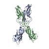
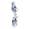
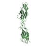
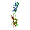
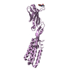
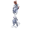
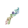
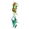
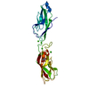
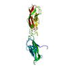
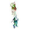
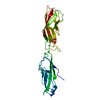
| Entry | Database: PDB / ID: 1ncj | ||||||
|---|---|---|---|---|---|---|---|
| Title | N-CADHERIN, TWO-DOMAIN FRAGMENT | ||||||
 Components Components | PROTEIN (N-CADHERIN) | ||||||
 Keywords Keywords |  CELL ADHESION PROTEIN CELL ADHESION PROTEIN | ||||||
| Function / homology |  Function and homology information Function and homology informationmesenchymal cell migration / regulation of oligodendrocyte progenitor proliferation / regulation of postsynaptic density protein 95 clustering / radial glial cell differentiation / neuroligin clustering involved in postsynaptic membrane assembly / positive regulation of synaptic vesicle clustering / Regulation of Insulin-like Growth Factor (IGF) transport and uptake by Insulin-like Growth Factor Binding Proteins (IGFBPs) / Post-translational protein phosphorylation / Adherens junctions interactions / desmosome ...mesenchymal cell migration / regulation of oligodendrocyte progenitor proliferation / regulation of postsynaptic density protein 95 clustering / radial glial cell differentiation / neuroligin clustering involved in postsynaptic membrane assembly / positive regulation of synaptic vesicle clustering / Regulation of Insulin-like Growth Factor (IGF) transport and uptake by Insulin-like Growth Factor Binding Proteins (IGFBPs) / Post-translational protein phosphorylation / Adherens junctions interactions / desmosome / synaptic vesicle clustering / gamma-catenin binding / neural crest cell development / glial cell differentiation / telencephalon development / neuroepithelial cell differentiation / type B pancreatic cell development / cell-cell adhesion mediated by cadherin / neuronal stem cell population maintenance / alpha-catenin binding / fascia adherens / apicolateral plasma membrane / calcium-dependent cell-cell adhesion via plasma membrane cell adhesion molecules / Myogenesis / regulation of Rho protein signal transduction / postsynaptic specialization membrane / brain morphogenesis / catenin complex / cell-cell junction assembly / adherens junction organization / blood vessel morphogenesis / regulation of myelination / heterophilic cell-cell adhesion via plasma membrane cell adhesion molecules / cortical actin cytoskeleton / regulation of axonogenesis / nitric-oxide synthase binding / homophilic cell adhesion via plasma membrane adhesion molecules / plasma membrane raft / intercalated disc / homeostasis of number of cells / presynaptic active zone membrane / regulation of synaptic transmission, glutamatergic / T-tubule / synapse assembly / striated muscle cell differentiation / protein tyrosine kinase binding / protein localization to plasma membrane / adherens junction / brain development / modulation of chemical synaptic transmission / negative regulation of canonical Wnt signaling pathway / cell morphogenesis / sarcolemma / cerebral cortex development / beta-catenin binding / cell-cell adhesion / regulation of protein localization / cell-cell junction / cell migration / apical part of cell / lamellipodium / basolateral plasma membrane / protein phosphatase binding / positive regulation of MAPK cascade / postsynaptic density / cell adhesion / neuron projection / cadherin binding / apical plasma membrane / glutamatergic synapse / synapse / calcium ion binding / protein-containing complex binding / protein kinase binding / enzyme binding / cell surface / protein-containing complex / RNA binding / membrane / identical protein binding / plasma membrane / cytoplasmSimilarity search - Function | ||||||
| Biological species |  Mus musculus (house mouse) Mus musculus (house mouse) | ||||||
| Method | X-RAY DIFFRACTION / SYNCHROTRON / MOLECULAR REPLACEMENT / Resolution: 3.4 Å | ||||||
 Authors Authors | Tamura, K. / Shan, W.-S. / Hendrickson, W.A. / Colman, D.R. / Shapiro, L. | ||||||
 Citation Citation | Journal: Neuron / Year: 1998 Title: Structure-function analysis of cell adhesion by neural (N-) cadherin. Authors: Tamura, K. / Shan, W.S. / Hendrickson, W.A. / Colman, D.R. / Shapiro, L. | ||||||
| History |
|
- Structure visualization
| Structure viewer | Molecule: MolmilJmol/JSmol |
|---|
- Downloads & links
-Download
| PDBx/mmCIF format | 1ncj.cif.gz | 60.3 KB | Display | PDBx/mmCIF format |
|---|---|---|---|---|
| PDB format | pdb1ncj.ent.gz | 43.3 KB | Display | PDB format |
| PDBx/mmJSON format | 1ncj.json.gz | Tree view | PDBx/mmJSON format | |
| Others |  Other downloads Other downloads |
-Validation report
| Arichive directory | https://data.pdbj.org/pub/pdb/validation_reports/nc/1ncjftp://data.pdbj.org/pub/pdb/validation_reports/nc/1ncj | HTTPS FTP |
|---|
-Related structure data


-Links
 PDBj
PDBj
- Assembly
| Deposited unit | 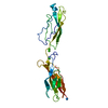
| ||||||||
|---|---|---|---|---|---|---|---|---|---|
| 1 |
| ||||||||
| Unit cell |
| ||||||||
| Components on special symmetry positions |
|
-Components
| #1: Protein | Mass: 23603.465 Da / Num. of mol.: 1 / Fragment: TWO-DOMAIN FRAGMENT Source method: isolated from a genetically manipulated source Source: (gene. exp.) Mus musculus (house mouse) / Tissue: BRAIN / Production host:  Escherichia coli (E. coli) / References: UniProt: P15116 Escherichia coli (E. coli) / References: UniProt: P15116 | ||
|---|---|---|---|
| #2: Chemical |   Mass: 40.078 Da / Num. of mol.: 3 / Source method: obtained synthetically / Formula: Ca Mass: 40.078 Da / Num. of mol.: 3 / Source method: obtained synthetically / Formula: Ca#3: Chemical | ChemComp-IUM / | Uranyl  Mass: 270.028 Da / Num. of mol.: 1 / Source method: obtained synthetically / Formula: O2U Mass: 270.028 Da / Num. of mol.: 1 / Source method: obtained synthetically / Formula: O2U |
-Experimental details
-Experiment
| Experiment | Method: X-RAY DIFFRACTION / Number of used crystals: 1 |
|---|
- Sample preparation
| Crystal | Density Matthews: 3.53 Å3/Da / Density % sol: 65.15 % | ||||||||||||||||||||||||||||||
|---|---|---|---|---|---|---|---|---|---|---|---|---|---|---|---|---|---|---|---|---|---|---|---|---|---|---|---|---|---|---|---|
| Crystal grow | pH: 5 Details: REMARK 280 CRYSTALLIZATION CONDITIONS: REMARK 280 HANGING DROP, 20MG/ML PROTEIN REMARK 280 50 MM NA ACETATE, PH 5.0, 15MM CACL2, REMARK 280 1MM UO2 ACETATE | ||||||||||||||||||||||||||||||
| Crystal grow | *PLUS Method: vapor diffusionDetails: drop contains equal volume of protein and reservoir solutions | ||||||||||||||||||||||||||||||
| Components of the solutions | *PLUS
|
-Data collection
| Diffraction | Mean temperature: 100 K |
|---|---|
| Diffraction source | Source: SYNCHROTRON / Site: NSLS  / Beamline: X4A / Wavelength: 0.97 / Beamline: X4A / Wavelength: 0.97 |
| Detector | Type: FUJI / Detector: IMAGE PLATE / Date: Jan 1, 1997 / Details: MIRRORS |
| Radiation | Monochromator: SI / Protocol: SINGLE WAVELENGTH / Monochromatic (M) / Laue (L): M / Scattering type: x-ray |
| Radiation wavelength | Wavelength: 0.97 Å / Relative weight: 1 |
| Reflection | Resolution: 3.4→20 Å / Num. obs: 9451 / % possible obs: 98.6 % / Observed criterion σ(I): 0 / Redundancy: 4 % / Rmerge(I) obs: 0.93 / Rsym value: 0.93 / Net I/σ(I): 12.96 |
| Reflection shell | Resolution: 3.4→3.5 Å / Redundancy: 3 % / Rmerge(I) obs: 0.322 / Mean I/σ(I) obs: 3.12 / Rsym value: 0.322 / % possible all: 97.7 |
| Reflection shell | *PLUS % possible obs: 97.7 % |
- Processing
| Software |
| ||||||||||||||||||||||||||||||||||||||||||||||||||||||||||||
|---|---|---|---|---|---|---|---|---|---|---|---|---|---|---|---|---|---|---|---|---|---|---|---|---|---|---|---|---|---|---|---|---|---|---|---|---|---|---|---|---|---|---|---|---|---|---|---|---|---|---|---|---|---|---|---|---|---|---|---|---|---|
| Refinement | Method to determine structure: MOLECULAR REPLACEMENT Starting model: 1NCG AND 1EDH Resolution: 3.4→6 Å / Cross valid method: THROUGHOUT / σ(F): 2
| ||||||||||||||||||||||||||||||||||||||||||||||||||||||||||||
| Displacement parameters | Biso mean: 29.2 Å2 | ||||||||||||||||||||||||||||||||||||||||||||||||||||||||||||
| Refinement step | Cycle: LAST / Resolution: 3.4→6 Å
| ||||||||||||||||||||||||||||||||||||||||||||||||||||||||||||
| Refine LS restraints |
| ||||||||||||||||||||||||||||||||||||||||||||||||||||||||||||
| LS refinement shell | Resolution: 3.4→3.55 Å / Total num. of bins used: 8 / % reflection obs: 97 % | ||||||||||||||||||||||||||||||||||||||||||||||||||||||||||||
| Xplor file | Serial no: 1 / Param file: PARAM19X.PRO / Topol file: TOPH19X.PRO | ||||||||||||||||||||||||||||||||||||||||||||||||||||||||||||
| Software | *PLUS Name: X-PLOR / Version: 3.1 / Classification: refinement | ||||||||||||||||||||||||||||||||||||||||||||||||||||||||||||
| Refinement | *PLUS Rfactor obs: 0.212 | ||||||||||||||||||||||||||||||||||||||||||||||||||||||||||||
| Solvent computation | *PLUS | ||||||||||||||||||||||||||||||||||||||||||||||||||||||||||||
| Displacement parameters | *PLUS | ||||||||||||||||||||||||||||||||||||||||||||||||||||||||||||
| Refine LS restraints | *PLUS
|
