 Movie
Movie Controller


+ Open data

- Basic information
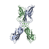
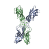
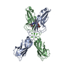
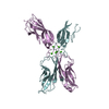
| Entry | Database: PDB / ID: 1edh | ||||||
|---|---|---|---|---|---|---|---|
| Title | E-CADHERIN DOMAINS 1 AND 2 IN COMPLEX WITH CALCIUM | ||||||
 Components Components | E-CADHERIN Cadherin-1 Cadherin-1 | ||||||
 Keywords Keywords | CELL ADHESION PROTEIN / CADHERIN / CALCIUM BINDING PROTEIN | ||||||
| Function / homology |  Function and homology information Function and homology informationuterine epithelium development / Apoptotic cleavage of cell adhesion proteins / salivary gland cavitation / regulation of branching involved in salivary gland morphogenesis / regulation of protein catabolic process at postsynapse, modulating synaptic transmission / RHO GTPases activate IQGAPs / Adherens junctions interactions / Degradation of the extracellular matrix / Integrin cell surface interactions / regulation of neuron migration ...uterine epithelium development / Apoptotic cleavage of cell adhesion proteins / salivary gland cavitation / regulation of branching involved in salivary gland morphogenesis / regulation of protein catabolic process at postsynapse, modulating synaptic transmission / RHO GTPases activate IQGAPs / Adherens junctions interactions / Degradation of the extracellular matrix / Integrin cell surface interactions / regulation of neuron migration / positive regulation of cell-cell adhesion / lateral loop / cell-cell adhesion mediated by cadherin / negative regulation of axon extension / regulation of protein localization to cell surface / trophectodermal cell differentiation / alpha-catenin binding / flotillin complex / epithelial cell morphogenesis / Schmidt-Lanterman incisure / bicellular tight junction assembly / calcium-dependent cell-cell adhesion via plasma membrane cell adhesion molecules / intestinal epithelial cell development / node of Ranvier / protein metabolic process / catenin complex / negative regulation of protein processing / cell-cell junction assembly / negative regulation of protein localization to plasma membrane / adherens junction organization / apical junction complex / cochlea development / homophilic cell adhesion via plasma membrane adhesion molecules / microvillus / decidualization / canonical Wnt signaling pathway / establishment of skin barrier / axon terminus / synapse assembly / cytoskeletal protein binding / embryo implantation / protein tyrosine kinase binding / protein localization to plasma membrane / cell periphery / sensory perception of sound / cellular response to amino acid stimulus / adherens junction / negative regulation of canonical Wnt signaling pathway / cell morphogenesis / beta-catenin binding / cell-cell adhesion / regulation of protein localization / negative regulation of epithelial cell proliferation / cell-cell junction / apical part of cell / actin cytoskeleton organization / postsynapse / regulation of gene expression / basolateral plasma membrane / protein phosphatase binding / in utero embryonic development / molecular adaptor activity / endosome / cadherin binding / protein domain specific binding / axon / glutamatergic synapse / calcium ion binding / Golgi apparatus / cell surface / identical protein binding / plasma membrane / cytoplasmSimilarity search - Function | ||||||
| Biological species |  Mus musculus (house mouse) Mus musculus (house mouse) | ||||||
| Method | X-RAY DIFFRACTION / SYNCHROTRON / THE INITIAL MODEL WAS OBTAINED FROM A COMBINATION OF MULTI-WAVELENGTH ANOMALOUS DIFFRACTION (MAD), REAL SPACE AVERAGING TECHNIQUES. THE MAD DATA WAS TAKEN AT THE MERCURY L(III) EDGE AT 3 DIFFERENT WAVELENGTHS (CHESS BEAMLINE F2). REAL SPACE AVERAGING WAS USED TO IMPROVE THE MAPS. / Resolution: 2 Å | ||||||
 Authors Authors | Nagar, B. / Overduin, M. / Ikura, M. / Rini, J.M. | ||||||
 Citation Citation | Journal: Nature / Year: 1996 Title: Structural basis of calcium-induced E-cadherin rigidification and dimerization. Authors: Nagar, B. / Overduin, M. / Ikura, M. / Rini, J.M. | ||||||
| History |
|
- Structure visualization
| Structure viewer | Molecule: MolmilJmol/JSmol |
|---|
- Downloads & links
-Download
| PDBx/mmCIF format | 1edh.cif.gz | 100.6 KB | Display | PDBx/mmCIF format |
|---|---|---|---|---|
| PDB format | pdb1edh.ent.gz | 75.5 KB | Display | PDB format |
| PDBx/mmJSON format | 1edh.json.gz | Tree view | PDBx/mmJSON format | |
| Others |  Other downloads Other downloads |
-Validation report
| Arichive directory | https://data.pdbj.org/pub/pdb/validation_reports/ed/1edhftp://data.pdbj.org/pub/pdb/validation_reports/ed/1edh | HTTPS FTP |
|---|
-Related structure data
| Similar structure data |
|---|


-Links
 PDBj
PDBj


- Assembly
| Deposited unit | 
| ||||||||
|---|---|---|---|---|---|---|---|---|---|
| 1 |
| ||||||||
| Unit cell |
| ||||||||
| Details | THE ASYMMETRIC UNIT (MOLECULES A AND B) CONTAINS A CALCIUM MEDIATED DIMER. BOTH MOLECULES IN THE ASYMMETRIC UNIT CONTAIN THREE CALCIUM IONS (CA 2+) AND ONE MERCURY ATOM (HG). THE MERCURY ATOMS ARE BOUND TO AN EXPOSED CYSTEINE RESIDUE IN THE N-TERMINAL DOMAIN. |
-Components
| #1: Protein | Cadherin-1 / EPITHELIAL CADHERIN DOMAINS 1 AND 2 / ECAD12 Mass: 24718.504 Da / Num. of mol.: 2 / Mutation: INS(MR-D1) Source method: isolated from a genetically manipulated source Source: (gene. exp.) Mus musculus (house mouse) / Tissue: EPITHELIAL / Cellular location: CELL SURFACECell membrane / Plasmid: PAS / Production host:  Escherichia coli (E. coli) / Strain (production host): AR58 / References: UniProt: P09803 Escherichia coli (E. coli) / Strain (production host): AR58 / References: UniProt: P09803#2: Chemical | Mercury (element)  Mass: 200.590 Da / Num. of mol.: 2 / Source method: obtained synthetically / Formula: Hg Mass: 200.590 Da / Num. of mol.: 2 / Source method: obtained synthetically / Formula: Hg#3: Chemical | ChemComp-CA /   Mass: 40.078 Da / Num. of mol.: 6 / Source method: obtained synthetically / Formula: Ca Mass: 40.078 Da / Num. of mol.: 6 / Source method: obtained synthetically / Formula: Ca#4: Water | ChemComp-HOH / | Water Mass: 18.015 Da / Num. of mol.: 249 / Source method: isolated from a natural source / Formula: H2O Mass: 18.015 Da / Num. of mol.: 249 / Source method: isolated from a natural source / Formula: H2O |
|---|
-Experimental details
-Experiment
| Experiment | Method: X-RAY DIFFRACTION / Number of used crystals: 1 |
|---|
- Sample preparation
| Crystal | Density Matthews: 3.23 Å3/Da / Density % sol: 62 % | |||||||||||||||||||||||||||||||||||||||||||||
|---|---|---|---|---|---|---|---|---|---|---|---|---|---|---|---|---|---|---|---|---|---|---|---|---|---|---|---|---|---|---|---|---|---|---|---|---|---|---|---|---|---|---|---|---|---|---|
| Crystal grow | pH: 9 Details: 1.2 M AMMONIUM SULFATE 0.01 M CALCIUM CHLORIDE 0.1 M TRIS-HCL BUFFER, PH 9.0 0.003 M SODIUM AZIDE | |||||||||||||||||||||||||||||||||||||||||||||
| Crystal grow | *PLUS pH: 7.25 / Method: vapor diffusion | |||||||||||||||||||||||||||||||||||||||||||||
| Components of the solutions | *PLUS
|
-Data collection
| Diffraction | Mean temperature: 113 K |
|---|---|
| Diffraction source | Source: SYNCHROTRON / Site: CHESS  / Beamline: A1 / Wavelength: 0.908 / Beamline: A1 / Wavelength: 0.908 |
| Detector | Type: PRINCETON 2K / Detector: CCD / Date: Apr 3, 1995 |
| Radiation | Monochromator: SI(111) / Monochromatic (M) / Laue (L): M / Scattering type: x-ray |
| Radiation wavelength | Wavelength: 0.908 Å / Relative weight: 1 |
| Reflection | Resolution: 2→22 Å / Num. obs: 37605 / % possible obs: 88.3 % / Observed criterion σ(I): 2 / Redundancy: 2.3 % / Biso Wilson estimate: 26.08 Å2 / Rsym value: 0.053 / Net I/σ(I): 13 |
| Reflection shell | Resolution: 2→2.07 Å / Redundancy: 1.59 % / Rsym value: 0.146 / % possible all: 63.6 |
| Reflection | *PLUS Num. measured all: 86202 / Rmerge(I) obs: 0.053 |
- Processing
| Software |
| ||||||||||||||||||||||||||||||||||||||||||||||||||||||||||||
|---|---|---|---|---|---|---|---|---|---|---|---|---|---|---|---|---|---|---|---|---|---|---|---|---|---|---|---|---|---|---|---|---|---|---|---|---|---|---|---|---|---|---|---|---|---|---|---|---|---|---|---|---|---|---|---|---|---|---|---|---|---|
| Refinement | Method to determine structure: THE INITIAL MODEL WAS OBTAINED FROM A COMBINATION OF MULTI-WAVELENGTH ANOMALOUS DIFFRACTION (MAD), REAL SPACE AVERAGING TECHNIQUES. THE MAD DATA WAS TAKEN AT THE ...Method to determine structure: THE INITIAL MODEL WAS OBTAINED FROM A COMBINATION OF MULTI-WAVELENGTH ANOMALOUS DIFFRACTION (MAD), REAL SPACE AVERAGING TECHNIQUES. THE MAD DATA WAS TAKEN AT THE MERCURY L(III) EDGE AT 3 DIFFERENT WAVELENGTHS (CHESS BEAMLINE F2). REAL SPACE AVERAGING WAS USED TO IMPROVE THE MAPS. Resolution: 2→5 Å / Data cutoff high absF: 83.4 / σ(F): 2 Details: THE ELECTRON DENSITY FOR THE LOOP BETWEEN STRANDS B AND C (RESIDUES 27 - 32) IS WEAK.
| ||||||||||||||||||||||||||||||||||||||||||||||||||||||||||||
| Displacement parameters | Biso mean: 29.89 Å2 | ||||||||||||||||||||||||||||||||||||||||||||||||||||||||||||
| Refinement step | Cycle: LAST / Resolution: 2→5 Å
| ||||||||||||||||||||||||||||||||||||||||||||||||||||||||||||
| Refine LS restraints |
| ||||||||||||||||||||||||||||||||||||||||||||||||||||||||||||
| LS refinement shell | Resolution: 2→2.07 Å
| ||||||||||||||||||||||||||||||||||||||||||||||||||||||||||||
| Xplor file |
| ||||||||||||||||||||||||||||||||||||||||||||||||||||||||||||
| Software | *PLUS Name: X-PLOR / Version: 3.1 / Classification: refinement | ||||||||||||||||||||||||||||||||||||||||||||||||||||||||||||
| Refine LS restraints | *PLUS
|
