 Movie
Movie Controller

[English] 日本語
 Yorodumi
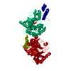
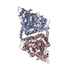
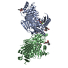
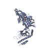
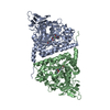
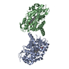
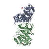
- PDB-1mzf: Human Factor inhibiting HIF (FIH1) in Complex with 2-oxoglutarate -
+ Open data

- Basic information
| Entry | Database: PDB / ID: 1mzf | ||||||
|---|---|---|---|---|---|---|---|
| Title | Human Factor inhibiting HIF (FIH1) in Complex with 2-oxoglutarate | ||||||
 Components Components | factor inhibiting HIF1 | ||||||
 Keywords Keywords |  OXIDOREDUCTASE / beta-jellyroll OXIDOREDUCTASE / beta-jellyroll | ||||||
| Function / homology |  Function and homology informationhypoxia-inducible factor-asparagine dioxygenase / : / [protein]-asparagine 3-dioxygenase activity / peptidyl-histidine dioxygenase activity / peptidyl-aspartic acid 3-dioxygenase activity / Cellular response to hypoxia / carboxylic acid binding / positive regulation of vasculogenesis / ankyrin repeat binding / oxygen sensor activity ...hypoxia-inducible factor-asparagine dioxygenase / : / [protein]-asparagine 3-dioxygenase activity / peptidyl-histidine dioxygenase activity / peptidyl-aspartic acid 3-dioxygenase activity / Cellular response to hypoxia / carboxylic acid binding / positive regulation of vasculogenesis / ankyrin repeat binding / oxygen sensor activity / Notch binding / negative regulation of Notch signaling pathway / NF-kappaB binding / positive regulation of myoblast differentiation / ferrous iron binding / transcription corepressor activity / perinuclear region of cytoplasm / protein homodimerization activity / zinc ion binding / nucleoplasm / cytosol / cytoplasm Function and homology informationhypoxia-inducible factor-asparagine dioxygenase / : / [protein]-asparagine 3-dioxygenase activity / peptidyl-histidine dioxygenase activity / peptidyl-aspartic acid 3-dioxygenase activity / Cellular response to hypoxia / carboxylic acid binding / positive regulation of vasculogenesis / ankyrin repeat binding / oxygen sensor activity ...hypoxia-inducible factor-asparagine dioxygenase / : / [protein]-asparagine 3-dioxygenase activity / peptidyl-histidine dioxygenase activity / peptidyl-aspartic acid 3-dioxygenase activity / Cellular response to hypoxia / carboxylic acid binding / positive regulation of vasculogenesis / ankyrin repeat binding / oxygen sensor activity / Notch binding / negative regulation of Notch signaling pathway / NF-kappaB binding / positive regulation of myoblast differentiation / ferrous iron binding / transcription corepressor activity / perinuclear region of cytoplasm / protein homodimerization activity / zinc ion binding / nucleoplasm / cytosol / cytoplasmSimilarity search - Function | ||||||
| Biological species |  Homo sapiens (human) Homo sapiens (human) | ||||||
| Method | X-RAY DIFFRACTION / SYNCHROTRON / MOLECULAR REPLACEMENT / Resolution: 2.4 Å | ||||||
 Authors Authors | Dann III, C.E. / Bruick, R.K. / Deisenhofer, J. | ||||||
 Citation Citation | Journal: Proc.Natl.Acad.Sci.USA / Year: 2002 Title: Structure of Factor-Inhibiting Hypoxia-Inducible Factor 1: An Asparaginyl Hydroxylase Involved in the Hypoxic Response Pathway. Authors: Dann III, C.E. / Bruick, R.K. / Deisenhofer, J. | ||||||
| History |
|
- Structure visualization
| Structure viewer | Molecule: MolmilJmol/JSmol |
|---|
- Downloads & links
-Download
| PDBx/mmCIF format | 1mzf.cif.gz | 82.9 KB | Display | PDBx/mmCIF format |
|---|---|---|---|---|
| PDB format | pdb1mzf.ent.gz | 61.9 KB | Display | PDB format |
| PDBx/mmJSON format | 1mzf.json.gz | Tree view | PDBx/mmJSON format | |
| Others |  Other downloads Other downloads |
-Validation report
| Arichive directory | https://data.pdbj.org/pub/pdb/validation_reports/mz/1mzfftp://data.pdbj.org/pub/pdb/validation_reports/mz/1mzf | HTTPS FTP |
|---|
-Related structure data


-Links
 PDBj
PDBj


- Assembly
| Deposited unit | 
| ||||||||
|---|---|---|---|---|---|---|---|---|---|
| 1 | 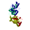
| ||||||||
| 2 | 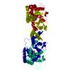
| ||||||||
| Unit cell |
|
-Components
| #1: Protein | Mass: 40456.406 Da / Num. of mol.: 1 Source method: isolated from a genetically manipulated source Source: (gene. exp.) Homo sapiens (human) / Gene: FIH-1 / Plasmid: pHis-parallel / Production host:  Escherichia coli (E. coli) / Strain (production host): Rosetta(DE3)pLysS / References: UniProt: Q9NWT6 Escherichia coli (E. coli) / Strain (production host): Rosetta(DE3)pLysS / References: UniProt: Q9NWT6 |
|---|---|
| #2: Chemical | ChemComp-FE2 /   Mass: 55.845 Da / Num. of mol.: 1 / Source method: obtained synthetically / Formula: Fe Mass: 55.845 Da / Num. of mol.: 1 / Source method: obtained synthetically / Formula: Fe |
| #3: Chemical | ChemComp-AKG / Α-Ketoglutaric acid  Mass: 146.098 Da / Num. of mol.: 1 / Source method: obtained synthetically / Formula: C5H6O5 Mass: 146.098 Da / Num. of mol.: 1 / Source method: obtained synthetically / Formula: C5H6O5 |
| #4: Water | ChemComp-HOH / Water Mass: 18.015 Da / Num. of mol.: 50 / Source method: isolated from a natural source / Formula: H2O Mass: 18.015 Da / Num. of mol.: 50 / Source method: isolated from a natural source / Formula: H2O |
-Experimental details
-Experiment
| Experiment | Method: X-RAY DIFFRACTION / Number of used crystals: 1 |
|---|
- Sample preparation
| Crystal | Density Matthews: 3.41 Å3/Da / Density % sol: 63.97 % | ||||||||||||||||||||||||||||||||||||||||||||||||||||||||||||||||||||||
|---|---|---|---|---|---|---|---|---|---|---|---|---|---|---|---|---|---|---|---|---|---|---|---|---|---|---|---|---|---|---|---|---|---|---|---|---|---|---|---|---|---|---|---|---|---|---|---|---|---|---|---|---|---|---|---|---|---|---|---|---|---|---|---|---|---|---|---|---|---|---|---|
| Crystal grow | Temperature: 294 K / Method: vapor diffusion, hanging drop / pH: 9.6 Details: potassium tartrate, CAPSO, iron(II) sulfate, 2-oxoglutarate, glycerol, pH 9.6, VAPOR DIFFUSION, HANGING DROP, temperature 294K | ||||||||||||||||||||||||||||||||||||||||||||||||||||||||||||||||||||||
| Crystal grow | *PLUS Temperature: 21 ℃ / pH: 8 | ||||||||||||||||||||||||||||||||||||||||||||||||||||||||||||||||||||||
| Components of the solutions | *PLUS
|
-Data collection
| Diffraction | Mean temperature: 100 K |
|---|---|
| Diffraction source | Source: SYNCHROTRON / Site: ALS  / Beamline: 8.2.2 / Wavelength: 1.0781 Å / Beamline: 8.2.2 / Wavelength: 1.0781 Å |
| Detector | Type: ADSC QUANTUM 315 / Detector: CCD / Date: Sep 18, 2002 |
| Radiation | Monochromator: Si grating / Protocol: SINGLE WAVELENGTH / Monochromatic (M) / Laue (L): M / Scattering type: x-ray |
| Radiation wavelength | Wavelength: 1.0781 Å / Relative weight: 1 |
| Reflection | Resolution: 2.4→47.11 Å / Num. all: 22616 / Num. obs: 21549 / % possible obs: 95.3 % / Observed criterion σ(F): 0 / Observed criterion σ(I): 0 / Biso Wilson estimate: 44 Å2 / Limit h max: 36 / Limit h min: 0 / Limit k max: 25 / Limit k min: 0 / Limit l max: 61 / Limit l min: 0 / Observed criterion F max: 527737.13 / Observed criterion F min: 0.32 / Rsym value: 0.076 / Net I/σ(I): 21.8 |
| Reflection shell | Resolution: 2.4→2.49 Å / Mean I/σ(I) obs: 5.1 / Num. unique all: 2194 / Rsym value: 0.437 / % possible all: 99.9 |
| Reflection | *PLUS Highest resolution: 2.4 Å / Lowest resolution: 50 Å / Num. obs: 21793 / % possible obs: 98.2 % / Num. measured all: 172309 / Rmerge(I) obs: 0.076 |
| Reflection shell | *PLUS % possible obs: 99.9 % / Rmerge(I) obs: 0.443 |
- Processing
| Software |
| ||||||||||||||||||||||||||||||||||||||||||||||||||||||||||||||||||||||||||||||||||||||||||
|---|---|---|---|---|---|---|---|---|---|---|---|---|---|---|---|---|---|---|---|---|---|---|---|---|---|---|---|---|---|---|---|---|---|---|---|---|---|---|---|---|---|---|---|---|---|---|---|---|---|---|---|---|---|---|---|---|---|---|---|---|---|---|---|---|---|---|---|---|---|---|---|---|---|---|---|---|---|---|---|---|---|---|---|---|---|---|---|---|---|---|---|
| Refinement | Method to determine structure: MOLECULAR REPLACEMENT Starting model: FIH-1 Resolution: 2.4→29.93 Å / Rfactor Rfree error: 0.008 / Occupancy max: 1 / Occupancy min: 0 / Cross valid method: THROUGHOUT / σ(F): 0 / Stereochemistry target values: Engh & Huber
| ||||||||||||||||||||||||||||||||||||||||||||||||||||||||||||||||||||||||||||||||||||||||||
| Solvent computation | Solvent model: CNS bulk solvent model used / Bsol: 51.8402 Å2 / ksol: 0.357607 e/Å3 | ||||||||||||||||||||||||||||||||||||||||||||||||||||||||||||||||||||||||||||||||||||||||||
| Displacement parameters | Biso max: 109.69 Å2 / Biso mean: 60.46 Å2 / Biso min: 19.17 Å2
| ||||||||||||||||||||||||||||||||||||||||||||||||||||||||||||||||||||||||||||||||||||||||||
| Refine analyze |
| ||||||||||||||||||||||||||||||||||||||||||||||||||||||||||||||||||||||||||||||||||||||||||
| Refinement step | Cycle: LAST / Resolution: 2.4→29.93 Å
| ||||||||||||||||||||||||||||||||||||||||||||||||||||||||||||||||||||||||||||||||||||||||||
| Refine LS restraints |
| ||||||||||||||||||||||||||||||||||||||||||||||||||||||||||||||||||||||||||||||||||||||||||
| LS refinement shell | Refine-ID: X-RAY DIFFRACTION / Total num. of bins used: 8
| ||||||||||||||||||||||||||||||||||||||||||||||||||||||||||||||||||||||||||||||||||||||||||
| Xplor file |
| ||||||||||||||||||||||||||||||||||||||||||||||||||||||||||||||||||||||||||||||||||||||||||
| Software | *PLUS Name: CNS / Classification: refinement | ||||||||||||||||||||||||||||||||||||||||||||||||||||||||||||||||||||||||||||||||||||||||||
| Refinement | *PLUS Highest resolution: 2.4 Å / Lowest resolution: 30 Å / % reflection Rfree: 5 % | ||||||||||||||||||||||||||||||||||||||||||||||||||||||||||||||||||||||||||||||||||||||||||
| Solvent computation | *PLUS | ||||||||||||||||||||||||||||||||||||||||||||||||||||||||||||||||||||||||||||||||||||||||||
| Displacement parameters | *PLUS | ||||||||||||||||||||||||||||||||||||||||||||||||||||||||||||||||||||||||||||||||||||||||||
| Refine LS restraints | *PLUS
|
