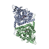
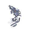
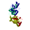
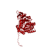
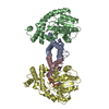
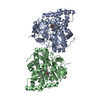
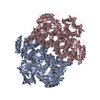
- PDB-5e5c: Crystal structure of dihydropyrimidinase from Pseudomonas aerugin... -
+
Open data
ID or keywords:
Loading...
-
Basic information
Entry
Database: PDB / ID: 5e5c
Title
Crystal structure of dihydropyrimidinase from Pseudomonas aeruginosa PAO1
Components
D-hydantoinase/dihydropyrimidinase
Keywords
HYDROLASE / dihydropyrimidinase
Function / homology
Function and homology information
dihydropyrimidinase / dihydropyrimidinase activity / hydrolase activity, acting on carbon-nitrogen (but not peptide) bonds, in cyclic amides / metal ion binding / cytosol Similarity search - Function
Resolution: 2.1→97.11 Å / Cor.coef. Fo:Fc: 0.961 / Cor.coef. Fo:Fc free: 0.934 / SU B: 4.515 / SU ML: 0.116 / Cross valid method: THROUGHOUT / ESU R: 0.172 / ESU R Free: 0.158 / Stereochemistry target values: MAXIMUM LIKELIHOOD / Details: HYDROGENS HAVE BEEN ADDED IN THE RIDING POSITIONS
Rfactor
Num. reflection
% reflection
Selection details
Rfree
0.21546
3471
5.1 %
RANDOM
Rwork
0.16957
-
-
-
obs
0.17191
65158
99.56 %
-
Solvent computation
Ion probe radii: 0.8 Å / Shrinkage radii: 0.8 Å / VDW probe radii: 1.2 Å / Solvent model: MASK
 Movie
Movie Controller

 Yorodumi
 Open data
 Basic information
 Components
 Keywords
 HYDROLASE /
  Function and homology information

 Authors
 Citation
 Structure visualization
 Downloads & links
 Other downloads


 PDBj
PDBj

 Assembly
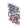

 Mass: 65.409 Da / Num. of mol.: 4 / Source method: obtained synthetically / Formula: Zn
 Mass: 18.015 Da / Num. of mol.: 338 / Source method: isolated from a natural source / Formula: H2O
 Sample preparation
 / Beamline: BL13C1 / Wavelength: 0.975 Å
 Processing