 Movie
Movie Controller


+ Open data

- Basic information
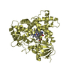
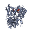
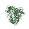
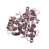
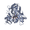
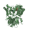
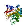
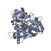
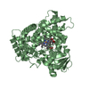
| Entry | Database: PDB / ID: 1mb9 | ||||||
|---|---|---|---|---|---|---|---|
| Title | BETA-LACTAM SYNTHETASE COMPLEXED WITH ATP | ||||||
 Components Components | BETA-LACTAM SYNTHETASE | ||||||
 Keywords Keywords |  HYDROLASE / CLAVULANIC ACID / ASPARAGINE SYNTHETASE / BETA-LACTAM SYNTHETASE / CARBOXYETHYL ARGININE / DEOXYGUANIDINOPROCLAVAMINIC ACID HYDROLASE / CLAVULANIC ACID / ASPARAGINE SYNTHETASE / BETA-LACTAM SYNTHETASE / CARBOXYETHYL ARGININE / DEOXYGUANIDINOPROCLAVAMINIC ACID | ||||||
| Function / homology |  Function and homology information(carboxyethyl)arginine beta-lactam-synthase / (carboxyethyl)arginine beta-lactam-synthase activity / asparagine synthase (glutamine-hydrolyzing) activity / asparagine biosynthetic process / clavulanic acid biosynthetic process / ATP binding / metal ion binding Function and homology information(carboxyethyl)arginine beta-lactam-synthase / (carboxyethyl)arginine beta-lactam-synthase activity / asparagine synthase (glutamine-hydrolyzing) activity / asparagine biosynthetic process / clavulanic acid biosynthetic process / ATP binding / metal ion bindingSimilarity search - Function | ||||||
| Biological species |  Streptomyces clavuligerus (bacteria) Streptomyces clavuligerus (bacteria) | ||||||
| Method | X-RAY DIFFRACTION / SYNCHROTRON / MOLECULAR REPLACEMENT / Resolution: 2.11 Å | ||||||
 Authors Authors | Miller, M.T. / Bachmann, B.O. / Townsend, C.A. / Rosenzweig, A.C. | ||||||
 Citation Citation | Journal: Proc.Natl.Acad.Sci.USA / Year: 2002 Title: The catalytic cycle of beta -lactam synthetase observed by x-ray crystallographic snapshots Authors: Miller, M.T. / Bachmann, B.O. / Townsend, C.A. / Rosenzweig, A.C. | ||||||
| History |
| ||||||
| Remark 600 | HETEROGEN ATP 701 HAS AN ALTERNATE CONFORMATION 'A'. AMP 706 AND POP 705 HAS ALTERNATE CONFORMATION 'B'. |
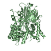
- Structure visualization
| Structure viewer | Molecule: MolmilJmol/JSmol |
|---|
- Downloads & links
-Download
| PDBx/mmCIF format | 1mb9.cif.gz | 209.7 KB | Display | PDBx/mmCIF format |
|---|---|---|---|---|
| PDB format | pdb1mb9.ent.gz | 165.2 KB | Display | PDB format |
| PDBx/mmJSON format | 1mb9.json.gz | Tree view | PDBx/mmJSON format | |
| Others |  Other downloads Other downloads |
-Validation report
| Arichive directory | https://data.pdbj.org/pub/pdb/validation_reports/mb/1mb9ftp://data.pdbj.org/pub/pdb/validation_reports/mb/1mb9 | HTTPS FTP |
|---|
-Related structure data
| Related structure data | 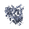 1m1zC  1mbzC 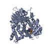 1mc1C 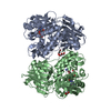 1jgtS S: Starting model for refinement C: citing same article ( |
|---|---|
| Similar structure data |
-Links
 PDBj
PDBj


- Assembly
| Deposited unit | 
| ||||||||
|---|---|---|---|---|---|---|---|---|---|
| 1 | 
| ||||||||
| 2 | 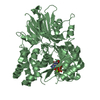
| ||||||||
| Unit cell |
|
-Components
-Protein , 1 types, 2 molecules AB
| #1: Protein | Mass: 54601.562 Da / Num. of mol.: 2 Source method: isolated from a genetically manipulated source Source: (gene. exp.) Streptomyces clavuligerus (bacteria) / Gene: 1901 / Plasmid: pET24a(+) / Species (production host): Escherichia coli / Production host: Escherichia coli BL21(DE3) (bacteria) / Strain (production host): BL21(DE3) / References: UniProt: Q9R8E3, UniProt: P0DJQ7*PLUS |
|---|
-Non-polymers , 5 types, 478 molecules 


| #2: Chemical | ChemComp-MG /  Mass: 24.305 Da / Num. of mol.: 4 / Source method: obtained synthetically / Formula: Mg Mass: 24.305 Da / Num. of mol.: 4 / Source method: obtained synthetically / Formula: Mg#3: Chemical | ChemComp-POP / | Pyrophosphate Mass: 175.959 Da / Num. of mol.: 1 / Source method: obtained synthetically / Formula: H2O7P2 Mass: 175.959 Da / Num. of mol.: 1 / Source method: obtained synthetically / Formula: H2O7P2#4: Chemical | ChemComp-AMP / | Adenosine monophosphate Mass: 347.221 Da / Num. of mol.: 1 / Source method: obtained synthetically / Formula: C10H14N5O7P / Comment: AMP*YM Mass: 347.221 Da / Num. of mol.: 1 / Source method: obtained synthetically / Formula: C10H14N5O7P / Comment: AMP*YM#5: Chemical | Adenosine triphosphate Mass: 507.181 Da / Num. of mol.: 2 / Source method: obtained synthetically / Formula: C10H16N5O13P3 / Comment: ATP, energy-carrying molecule*YM Mass: 507.181 Da / Num. of mol.: 2 / Source method: obtained synthetically / Formula: C10H16N5O13P3 / Comment: ATP, energy-carrying molecule*YM#6: Water | ChemComp-HOH / | WaterMass: 18.015 Da / Num. of mol.: 470 / Source method: isolated from a natural source / Formula: H2O |
|---|
-Experimental details
-Experiment
| Experiment | Method: X-RAY DIFFRACTION / Number of used crystals: 1 |
|---|
- Sample preparation
| Crystal | Density Matthews: 2.22 Å3/Da / Density % sol: 44.68 % | |||||||||||||||||||||||||||||||||||||||||||||||||
|---|---|---|---|---|---|---|---|---|---|---|---|---|---|---|---|---|---|---|---|---|---|---|---|---|---|---|---|---|---|---|---|---|---|---|---|---|---|---|---|---|---|---|---|---|---|---|---|---|---|---|
| Crystal grow | Temperature: 296 K / Method: vapor diffusion, sitting drop / pH: 8 Details: PEG 4000, Magnesium Chloride, Tris, pH 8.0, VAPOR DIFFUSION, SITTING DROP, temperature 296K | |||||||||||||||||||||||||||||||||||||||||||||||||
| Crystal grow | *PLUS Method: vapor diffusion, hanging drop / Details: Miller, M.T., (2001) Nature Struct. Biol., 8, 684. | |||||||||||||||||||||||||||||||||||||||||||||||||
| Components of the solutions | *PLUS
|
-Data collection
| Diffraction | Mean temperature: 100 K |
|---|---|
| Diffraction source | Source: SYNCHROTRON / Site: SSRL  / Beamline: BL9-1 / Wavelength: 1 Å / Beamline: BL9-1 / Wavelength: 1 Å |
| Detector | Type: MAR scanner 345 mm plate / Detector: IMAGE PLATE / Date: May 21, 2000 |
| Radiation | Monochromator: Curved Crystal / Protocol: SINGLE WAVELENGTH / Monochromatic (M) / Laue (L): M / Scattering type: x-ray |
| Radiation wavelength | Wavelength: 1 Å / Relative weight: 1 |
| Reflection | Resolution: 2.11→25 Å / Num. all: 64591 / Num. obs: 54280 / % possible obs: 99.6 % / Observed criterion σ(I): 5 / Biso Wilson estimate: 7.1 Å2 / Rsym value: 0.084 |
| Reflection shell | Resolution: 2.11→2.19 Å / Rsym value: 0.296 / % possible all: 98.5 |
| Reflection | *PLUS Highest resolution: 2.1 Å / Lowest resolution: 25 Å / Num. measured all: 594199 / Rmerge(I) obs: 0.084 |
| Reflection shell | *PLUS % possible obs: 98.5 % / Rmerge(I) obs: 0.296 |
- Processing
| Software |
| ||||||||||||||||||||||||||||||||||||
|---|---|---|---|---|---|---|---|---|---|---|---|---|---|---|---|---|---|---|---|---|---|---|---|---|---|---|---|---|---|---|---|---|---|---|---|---|---|
| Refinement | Method to determine structure: MOLECULAR REPLACEMENT Starting model: PDB ENTRY 1JGT Resolution: 2.11→24.78 Å / Rfactor Rfree error: 0.004 / Isotropic thermal model: RESTRAINED / Cross valid method: THROUGHOUT / σ(F): -3 / Stereochemistry target values: Engh & Huber
| ||||||||||||||||||||||||||||||||||||
| Solvent computation | Solvent model: FLAT MODEL / Bsol: 51.97 Å2 / ksol: 0.378188 e/Å3 | ||||||||||||||||||||||||||||||||||||
| Displacement parameters | Biso mean: 22.7 Å2
| ||||||||||||||||||||||||||||||||||||
| Refine analyze | Luzzati coordinate error free: 0.3 Å / Luzzati sigma a free: 0.24 Å | ||||||||||||||||||||||||||||||||||||
| Refinement step | Cycle: LAST / Resolution: 2.11→24.78 Å
| ||||||||||||||||||||||||||||||||||||
| Refine LS restraints |
| ||||||||||||||||||||||||||||||||||||
| LS refinement shell | Resolution: 2.11→2.24 Å / Rfactor Rfree error: 0.011 / Total num. of bins used: 6
| ||||||||||||||||||||||||||||||||||||
| Xplor file |
| ||||||||||||||||||||||||||||||||||||
| Refinement | *PLUS Lowest resolution: 25 Å / % reflection Rfree: 7.8 % / Rfactor Rwork: 0.21 | ||||||||||||||||||||||||||||||||||||
| Solvent computation | *PLUS | ||||||||||||||||||||||||||||||||||||
| Displacement parameters | *PLUS | ||||||||||||||||||||||||||||||||||||
| Refine LS restraints | *PLUS
|
