 Movie
Movie Controller


+ Open data

- Basic information
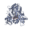
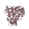
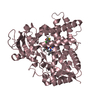
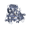
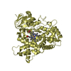
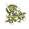
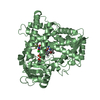
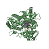
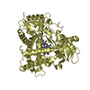
| Entry | Database: PDB / ID: 3t3r | ||||||
|---|---|---|---|---|---|---|---|
| Title | Human Cytochrome P450 2A6 in complex with Pilocarpine | ||||||
 Components Components | Cytochrome P450 2A6 | ||||||
 Keywords Keywords |  OXIDOREDUCTASE / CYP2A6 / cytochrome P450 2A6 / P450 2A6 / heme protein / monooxygenase / drug metabolism / xenobiotic metabolism / endoplasmic reticulum / membrane OXIDOREDUCTASE / CYP2A6 / cytochrome P450 2A6 / P450 2A6 / heme protein / monooxygenase / drug metabolism / xenobiotic metabolism / endoplasmic reticulum / membrane | ||||||
| Function / homology |  Function and homology information Function and homology informationcoumarin catabolic process / coumarin 7-hydroxylase activity / coumarin metabolic process / arachidonic acid epoxygenase activity / Oxidoreductases; Acting on paired donors, with incorporation or reduction of molecular oxygen; With reduced flavin or flavoprotein as one donor, and incorporation of one atom of oxygen into the other donor / CYP2E1 reactions / epoxygenase P450 pathway / Xenobiotics / oxidoreductase activity, acting on paired donors, with incorporation or reduction of molecular oxygen, reduced flavin or flavoprotein as one donor, and incorporation of one atom of oxygen / cytoplasmic microtubule ...coumarin catabolic process / coumarin 7-hydroxylase activity / coumarin metabolic process / arachidonic acid epoxygenase activity / Oxidoreductases; Acting on paired donors, with incorporation or reduction of molecular oxygen; With reduced flavin or flavoprotein as one donor, and incorporation of one atom of oxygen into the other donor / CYP2E1 reactions / epoxygenase P450 pathway / Xenobiotics / oxidoreductase activity, acting on paired donors, with incorporation or reduction of molecular oxygen, reduced flavin or flavoprotein as one donor, and incorporation of one atom of oxygen / cytoplasmic microtubule / steroid metabolic process / xenobiotic catabolic process / xenobiotic metabolic process / iron ion binding / intracellular membrane-bounded organelle / heme binding / endoplasmic reticulum membrane / enzyme binding / cytoplasmSimilarity search - Function | ||||||
| Biological species |  Homo sapiens (human) Homo sapiens (human) | ||||||
| Method | X-RAY DIFFRACTION / SYNCHROTRON / MOLECULAR REPLACEMENT / Resolution: 2.4 Å | ||||||
 Authors Authors | DeVore, N.M. / Scott, E.E. | ||||||
 Citation Citation | Journal: Febs J. / Year: 2012 Title: Structural comparison of cytochromes P450 2A6, 2A13, and 2E1 with pilocarpine. Authors: DeVore, N.M. / Meneely, K.M. / Bart, A.G. / Stephens, E.S. / Battaile, K.P. / Scott, E.E. | ||||||
| History |
|
- Structure visualization
| Structure viewer | Molecule: MolmilJmol/JSmol |
|---|
- Downloads & links
-Download
| PDBx/mmCIF format | 3t3r.cif.gz | 379.2 KB | Display | PDBx/mmCIF format |
|---|---|---|---|---|
| PDB format | pdb3t3r.ent.gz | 311.8 KB | Display | PDB format |
| PDBx/mmJSON format | 3t3r.json.gz | Tree view | PDBx/mmJSON format | |
| Others |  Other downloads Other downloads |
-Validation report
| Arichive directory | https://data.pdbj.org/pub/pdb/validation_reports/t3/3t3rftp://data.pdbj.org/pub/pdb/validation_reports/t3/3t3r | HTTPS FTP |
|---|
-Related structure data
| Related structure data |  3t3qC  3t3sC 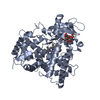 3t3zC  1z10S S: Starting model for refinement C: citing same article ( |
|---|---|
| Similar structure data |


-Links
 PDBj
PDBj

- Assembly
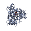
| Deposited unit | 
| ||||||||
|---|---|---|---|---|---|---|---|---|---|
| 1 | 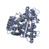
| ||||||||
| 2 | 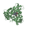
| ||||||||
| 3 | 
| ||||||||
| 4 | 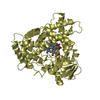
| ||||||||
| Unit cell |
|
-Components
| #1: Protein | Mass: 54671.637 Da / Num. of mol.: 4 / Fragment: unp residues 29-494 / Mutation: Y392F Source method: isolated from a genetically manipulated source Source: (gene. exp.) Homo sapiens (human) / Gene: CYP2A3, CYP2A6 / Plasmid: pKK2A6dH / Production host:  Escherichia coli (E. coli) / Strain (production host): TOPP3 / References: UniProt: P11509, unspecific monooxygenase Escherichia coli (E. coli) / Strain (production host): TOPP3 / References: UniProt: P11509, unspecific monooxygenase#2: Chemical | ChemComp-HEM / Heme B  Mass: 616.487 Da / Num. of mol.: 4 / Source method: obtained synthetically / Formula: C34H32FeN4O4 Mass: 616.487 Da / Num. of mol.: 4 / Source method: obtained synthetically / Formula: C34H32FeN4O4#3: Chemical | ChemComp-9PL / ( Pilocarpine  Mass: 208.257 Da / Num. of mol.: 4 / Source method: obtained synthetically / Formula: C11H16N2O2 / Comment: medication*YM Mass: 208.257 Da / Num. of mol.: 4 / Source method: obtained synthetically / Formula: C11H16N2O2 / Comment: medication*YM#4: Water | ChemComp-HOH / | Water Mass: 18.015 Da / Num. of mol.: 217 / Source method: isolated from a natural source / Formula: H2O Mass: 18.015 Da / Num. of mol.: 217 / Source method: isolated from a natural source / Formula: H2OSequence details | AUTHORS STATE THAT THE AMINOACID AT POSITION 392 OF THE UNIPROT ENTRY P11509 SHOULD BE A TYR | |
|---|
-Experimental details
-Experiment
| Experiment | Method: X-RAY DIFFRACTION / Number of used crystals: 1 |
|---|
- Sample preparation
| Crystal | Density Matthews: 2.65 Å3/Da / Density % sol: 53.62 % |
|---|---|
| Crystal grow | Temperature: 298 K / Method: vapor diffusion, hanging drop / pH: 8.5 Details: 30% PEG 3350, 0.175 M Tris, pH 8.5, 0.20 M ammomium sulfate, VAPOR DIFFUSION, HANGING DROP, temperature 298K |
-Data collection
| Diffraction | Mean temperature: 100 K |
|---|---|
| Diffraction source | Source: SYNCHROTRON / Site: SSRL  / Beamline: BL9-2 / Wavelength: 0.98 Å / Beamline: BL9-2 / Wavelength: 0.98 Å |
| Detector | Type: MARMOSAIC 325 mm CCD / Detector: CCD / Date: Jun 28, 2008 / Details: Rh coated flat mirror, toroidal focusing mirror |
| Radiation | Monochromator: Double crystal monochromator / Protocol: SINGLE WAVELENGTH / Monochromatic (M) / Laue (L): M / Scattering type: x-ray |
| Radiation wavelength | Wavelength: 0.98 Å / Relative weight: 1 |
| Reflection | Resolution: 2.4→87.04 Å / Num. all: 237442 / Num. obs: 85624 / % possible obs: 96.5 % / Observed criterion σ(F): 0 / Observed criterion σ(I): 0 / Redundancy: 2.8 % / Biso Wilson estimate: 41.4 Å2 / Rmerge(I) obs: 0.075 / Rsym value: 0.106 / Net I/σ(I): 13.2 |
| Reflection shell | Resolution: 2.4→2.46 Å / Redundancy: 2.7 % / Rmerge(I) obs: 0.402 / Mean I/σ(I) obs: 3.4 / Num. unique all: 17456 / % possible all: 97.9 |
- Processing
| Software |
| |||||||||||||||||||||||||||||||||||||||||||||||||||||||||||||||||
|---|---|---|---|---|---|---|---|---|---|---|---|---|---|---|---|---|---|---|---|---|---|---|---|---|---|---|---|---|---|---|---|---|---|---|---|---|---|---|---|---|---|---|---|---|---|---|---|---|---|---|---|---|---|---|---|---|---|---|---|---|---|---|---|---|---|---|
| Refinement | Method to determine structure: MOLECULAR REPLACEMENT Starting model: CYP2A6 PDB entry 1Z10 Resolution: 2.4→79 Å / Cor.coef. Fo:Fc: 0.935 / Cor.coef. Fo:Fc free: 0.893 / SU B: 8.008 / SU ML: 0.191 / Cross valid method: THROUGHOUT / σ(F): 0 / ESU R Free: 0.281 Stereochemistry target values: MAXIMUM LIKELIHOOD WITH PHASES
| |||||||||||||||||||||||||||||||||||||||||||||||||||||||||||||||||
| Solvent computation | Ion probe radii: 0.8 Å / Shrinkage radii: 0.8 Å / VDW probe radii: 1.4 Å / Solvent model: MASK | |||||||||||||||||||||||||||||||||||||||||||||||||||||||||||||||||
| Displacement parameters | Biso mean: 30.545 Å2
| |||||||||||||||||||||||||||||||||||||||||||||||||||||||||||||||||
| Refine analyze | Luzzati coordinate error obs: 0.277 Å | |||||||||||||||||||||||||||||||||||||||||||||||||||||||||||||||||
| Refinement step | Cycle: LAST / Resolution: 2.4→79 Å
| |||||||||||||||||||||||||||||||||||||||||||||||||||||||||||||||||
| Refine LS restraints |
| |||||||||||||||||||||||||||||||||||||||||||||||||||||||||||||||||
| LS refinement shell | Resolution: 2.4→2.462 Å / Total num. of bins used: 20
|
