 Movie
Movie Controller


+ Open data

- Basic information
| Entry | Database: PDB / ID: 1jgt | ||||||
|---|---|---|---|---|---|---|---|
| Title | CRYSTAL STRUCTURE OF BETA-LACTAM SYNTHETASE | ||||||
 Components Components | BETA-LACTAM SYNTHETASE | ||||||
 Keywords Keywords |  HYDROLASE / BETA-LACTAM SYNTHETASE / ASPARAGINE SYNTHETASE / CLAVULANIC ACID / AMPCPP / CEA / CARBOXYETHYLARGININE HYDROLASE / BETA-LACTAM SYNTHETASE / ASPARAGINE SYNTHETASE / CLAVULANIC ACID / AMPCPP / CEA / CARBOXYETHYLARGININE | ||||||
| Function / homology |  Function and homology information(carboxyethyl)arginine beta-lactam-synthase / (carboxyethyl)arginine beta-lactam-synthase activity / asparagine synthase (glutamine-hydrolyzing) activity / asparagine biosynthetic process / clavulanic acid biosynthetic process / ATP binding / metal ion binding Function and homology information(carboxyethyl)arginine beta-lactam-synthase / (carboxyethyl)arginine beta-lactam-synthase activity / asparagine synthase (glutamine-hydrolyzing) activity / asparagine biosynthetic process / clavulanic acid biosynthetic process / ATP binding / metal ion bindingSimilarity search - Function | ||||||
| Biological species |  Streptomyces clavuligerus (bacteria) Streptomyces clavuligerus (bacteria) | ||||||
| Method | X-RAY DIFFRACTION / SYNCHROTRON / MAD / Resolution: 1.95 Å | ||||||
 Authors Authors | Miller, M.T. / Bachmann, B.O. / Townsend, C.A. / Rosenzweig, A.C. | ||||||
 Citation Citation | Journal: Nat.Struct.Biol. / Year: 2001 Title: Structure of beta-lactam synthetase reveals how to synthesize antibiotics instead of asparagine. Authors: Miller, M.T. / Bachmann, B.O. / Townsend, C.A. / Rosenzweig, A.C. | ||||||
| History |
|
- Structure visualization
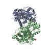
| Structure viewer | Molecule: MolmilJmol/JSmol |
|---|
- Downloads & links
-Download
| PDBx/mmCIF format | 1jgt.cif.gz | 216.2 KB | Display | PDBx/mmCIF format |
|---|---|---|---|---|
| PDB format | pdb1jgt.ent.gz | 168.5 KB | Display | PDB format |
| PDBx/mmJSON format | 1jgt.json.gz | Tree view | PDBx/mmJSON format | |
| Others |  Other downloads Other downloads |
-Validation report
| Arichive directory | https://data.pdbj.org/pub/pdb/validation_reports/jg/1jgtftp://data.pdbj.org/pub/pdb/validation_reports/jg/1jgt | HTTPS FTP |
|---|
-Related structure data
| Similar structure data |
|---|


-Links
 PDBj
PDBj


- Assembly
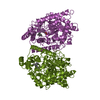
| Deposited unit | 
| ||||||||
|---|---|---|---|---|---|---|---|---|---|
| 1 |
| ||||||||
| Unit cell |
|
-Components
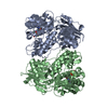
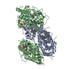
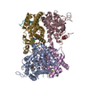
-Protein , 1 types, 2 molecules AB
| #1: Protein | Mass: 54601.562 Da / Num. of mol.: 2 Source method: isolated from a genetically manipulated source Source: (gene. exp.) Streptomyces clavuligerus (bacteria) / Gene: 1901 / Plasmid: pET24a(+) / Species (production host): Escherichia coli / Production host: Escherichia coli BL21(DE3) (bacteria) / Strain (production host): bl21(DE3) / References: UniProt: Q9R8E3, UniProt: P0DJQ7*PLUS |
|---|
-Non-polymers , 5 types, 633 molecules 


| #2: Chemical |  Mass: 24.305 Da / Num. of mol.: 2 / Source method: obtained synthetically / Formula: Mg Mass: 24.305 Da / Num. of mol.: 2 / Source method: obtained synthetically / Formula: Mg#3: Chemical |  Mass: 505.208 Da / Num. of mol.: 2 / Source method: obtained synthetically / Formula: C11H18N5O12P3 / Comment: AMP-CPP, energy-carrying molecule analogue*YM Mass: 505.208 Da / Num. of mol.: 2 / Source method: obtained synthetically / Formula: C11H18N5O12P3 / Comment: AMP-CPP, energy-carrying molecule analogue*YM#4: Chemical |  Mass: 246.264 Da / Num. of mol.: 2 / Source method: obtained synthetically / Formula: C9H18N4O4 Mass: 246.264 Da / Num. of mol.: 2 / Source method: obtained synthetically / Formula: C9H18N4O4#5: Chemical | ChemComp-GOL / Glycerol Mass: 92.094 Da / Num. of mol.: 5 / Source method: obtained synthetically / Formula: C3H8O3 Mass: 92.094 Da / Num. of mol.: 5 / Source method: obtained synthetically / Formula: C3H8O3#6: Water | ChemComp-HOH / | WaterMass: 18.015 Da / Num. of mol.: 622 / Source method: isolated from a natural source / Formula: H2O |
|---|
-Experimental details
-Experiment
| Experiment | Method: X-RAY DIFFRACTION / Number of used crystals: 1 |
|---|
- Sample preparation
| Crystal | Density Matthews: 2.22 Å3/Da / Density % sol: 44.71 % | |||||||||||||||||||||||||||||||||||||||||||||||||
|---|---|---|---|---|---|---|---|---|---|---|---|---|---|---|---|---|---|---|---|---|---|---|---|---|---|---|---|---|---|---|---|---|---|---|---|---|---|---|---|---|---|---|---|---|---|---|---|---|---|---|
| Crystal grow | Temperature: 296 K / Method: vapor diffusion, hanging drop / pH: 8 Details: POLYETHYLENEGLYCOL 4000, GLYCEROL, TRIS, pH 8.0, VAPOR DIFFUSION, HANGING DROP, temperature 296K | |||||||||||||||||||||||||||||||||||||||||||||||||
| Crystal grow | *PLUS pH: 7.5 / Details: Miller, M.T., (2001) Nature Struct. Biol., 8, 684. | |||||||||||||||||||||||||||||||||||||||||||||||||
| Components of the solutions | *PLUS
|
-Data collection
| Diffraction | Mean temperature: 100 K |
|---|---|
| Diffraction source | Source: SYNCHROTRON / Site: APS  / Beamline: 5ID-B / Wavelength: 1 Å / Beamline: 5ID-B / Wavelength: 1 Å |
| Detector | Type: MARRESEARCH / Detector: CCD / Date: Nov 18, 2000 |
| Radiation | Monochromator: GRAPHITE / Protocol: MAD / Monochromatic (M) / Laue (L): M / Scattering type: x-ray |
| Radiation wavelength | Wavelength: 1 Å / Relative weight: 1 |
| Reflection | Resolution: 1.95→19.97 Å / Num. all: 69641 / Num. obs: 68300 / % possible obs: 94.9 % / Observed criterion σ(F): 0 / Observed criterion σ(I): 1 / Redundancy: 6.8 % / Biso Wilson estimate: 15.1 Å2 / Limit h max: 31 / Limit h min: -31 / Limit k max: 50 / Limit k min: -31 / Limit l max: 41 / Limit l min: 0 / Observed criterion F max: 569036.55 / Observed criterion F min: 0.32 / Rmerge(I) obs: 0.194 / Net I/σ(I): 10.4 |
| Reflection shell | Resolution: 1.95→2.02 Å / Redundancy: 6.3 % / Rmerge(I) obs: 0.23 / % possible all: 98.1 |
| Reflection | *PLUS Lowest resolution: 20 Å / % possible obs: 98.3 % / Num. measured all: 465261 / Rmerge(I) obs: 0.062 |
| Reflection shell | *PLUS % possible obs: 98.1 % / Rmerge(I) obs: 0.231 |
- Processing
| Software |
| ||||||||||||||||||||||||||||||||||||||||||||||||||||||||||||||||||||||||||||||||||||||||||
|---|---|---|---|---|---|---|---|---|---|---|---|---|---|---|---|---|---|---|---|---|---|---|---|---|---|---|---|---|---|---|---|---|---|---|---|---|---|---|---|---|---|---|---|---|---|---|---|---|---|---|---|---|---|---|---|---|---|---|---|---|---|---|---|---|---|---|---|---|---|---|---|---|---|---|---|---|---|---|---|---|---|---|---|---|---|---|---|---|---|---|---|
| Refinement | Method to determine structure: MAD / Resolution: 1.95→19.89 Å / Rfactor Rfree error: 0.003 / Occupancy max: 1 / Occupancy min: 1 / Cross valid method: THROUGHOUT / σ(F): 0 / σ(I): 2 / Stereochemistry target values: Engh & Huber
| ||||||||||||||||||||||||||||||||||||||||||||||||||||||||||||||||||||||||||||||||||||||||||
| Solvent computation | Solvent model: CNS bulk solvent model used / Bsol: 55.1407 Å2 / ksol: 0.386944 e/Å3 | ||||||||||||||||||||||||||||||||||||||||||||||||||||||||||||||||||||||||||||||||||||||||||
| Displacement parameters | Biso max: 77.68 Å2 / Biso mean: 30.4 Å2 / Biso min: 11.7 Å2
| ||||||||||||||||||||||||||||||||||||||||||||||||||||||||||||||||||||||||||||||||||||||||||
| Refine analyze |
| ||||||||||||||||||||||||||||||||||||||||||||||||||||||||||||||||||||||||||||||||||||||||||
| Refinement step | Cycle: LAST / Resolution: 1.95→19.89 Å
| ||||||||||||||||||||||||||||||||||||||||||||||||||||||||||||||||||||||||||||||||||||||||||
| Refine LS restraints |
| ||||||||||||||||||||||||||||||||||||||||||||||||||||||||||||||||||||||||||||||||||||||||||
| LS refinement shell | Refine-ID: X-RAY DIFFRACTION / Total num. of bins used: 8
| ||||||||||||||||||||||||||||||||||||||||||||||||||||||||||||||||||||||||||||||||||||||||||
| Xplor file |
| ||||||||||||||||||||||||||||||||||||||||||||||||||||||||||||||||||||||||||||||||||||||||||
| Software | *PLUS Name: CNS / Version: 1 / Classification: refinement | ||||||||||||||||||||||||||||||||||||||||||||||||||||||||||||||||||||||||||||||||||||||||||
| Refinement | *PLUS Lowest resolution: 20 Å / σ(F): 0 | ||||||||||||||||||||||||||||||||||||||||||||||||||||||||||||||||||||||||||||||||||||||||||
| Solvent computation | *PLUS | ||||||||||||||||||||||||||||||||||||||||||||||||||||||||||||||||||||||||||||||||||||||||||
| Displacement parameters | *PLUS Biso mean: 30.4 Å2 | ||||||||||||||||||||||||||||||||||||||||||||||||||||||||||||||||||||||||||||||||||||||||||
| Refine LS restraints | *PLUS
| ||||||||||||||||||||||||||||||||||||||||||||||||||||||||||||||||||||||||||||||||||||||||||
| LS refinement shell | *PLUS Rfactor Rfree: 0.254 / % reflection Rfree: 7 % / Rfactor Rwork: 0.253 |
