 Movie
Movie Controller

[English] 日本語
 Yorodumi
- PDB-1ltj: Crystal Structure of Recombinant Human Fibrinogen Fragment D with... -
+ Open data

- Basic information
| Entry | Database: PDB / ID: 1ltj | |||||||||
|---|---|---|---|---|---|---|---|---|---|---|
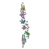
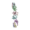
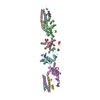
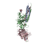
| Title | Crystal Structure of Recombinant Human Fibrinogen Fragment D with the Peptide Ligands Gly-Pro-Arg-Pro-Amide and Gly-His-Arg-Pro-Amide | |||||||||
 Components Components |
| |||||||||
 Keywords Keywords |  BLOOD CLOTTING / blood coagulation / fibrinogen / fibrinogen fragment D / recombinant fibrinogen fragment D / recombinant fibrinogen / recombinant fibrinogen fragment D with two peptide ligands BLOOD CLOTTING / blood coagulation / fibrinogen / fibrinogen fragment D / recombinant fibrinogen fragment D / recombinant fibrinogen / recombinant fibrinogen fragment D with two peptide ligands | |||||||||
| Function / homology |  Function and homology information Function and homology informationplatelet maturation / blood coagulation, common pathway / induction of bacterial agglutination / fibrinogen complex / Regulation of TLR by endogenous ligand / blood coagulation, fibrin clot formation / platelet alpha granule / cellular response to leptin stimulus / cellular response to interleukin-6 / MyD88 deficiency (TLR2/4) ...platelet maturation / blood coagulation, common pathway / induction of bacterial agglutination / fibrinogen complex / Regulation of TLR by endogenous ligand / blood coagulation, fibrin clot formation / platelet alpha granule / cellular response to leptin stimulus / cellular response to interleukin-6 / MyD88 deficiency (TLR2/4) / positive regulation of heterotypic cell-cell adhesion / IRAK4 deficiency (TLR2/4) / MyD88:MAL(TIRAP) cascade initiated on plasma membrane / extracellular matrix structural constituent / plasminogen activation / p130Cas linkage to MAPK signaling for integrins / positive regulation of exocytosis / positive regulation of peptide hormone secretion / GRB2:SOS provides linkage to MAPK signaling for Integrins / protein secretion / protein polymerization / Integrin cell surface interactions / cellular response to interleukin-1 / negative regulation of endothelial cell apoptotic process / Common Pathway of Fibrin Clot Formation / positive regulation of substrate adhesion-dependent cell spreading / negative regulation of extrinsic apoptotic signaling pathway via death domain receptors / positive regulation of vasoconstriction / fibrinolysis / cell adhesion molecule binding / Integrin signaling / cell-matrix adhesion / platelet alpha granule lumen / positive regulation of protein secretion / Post-translational protein phosphorylation / Signaling by high-kinase activity BRAF mutants / MAP2K and MAPK activation / platelet aggregation / response to calcium ion / Signaling by RAF1 mutants / Signaling by moderate kinase activity BRAF mutants / Paradoxical activation of RAF signaling by kinase inactive BRAF / Signaling downstream of RAS mutants / Regulation of Insulin-like Growth Factor (IGF) transport and uptake by Insulin-like Growth Factor Binding Proteins (IGFBPs) / extracellular vesicle / Signaling by BRAF and RAF1 fusions / Platelet degranulation / cell cortex / ER-Phagosome pathway / protein-folding chaperone binding / protein-containing complex assembly / collagen-containing extracellular matrix / blood microparticle / adaptive immune response / positive regulation of ERK1 and ERK2 cascade / Amyloid fiber formation / external side of plasma membrane / endoplasmic reticulum lumen / signaling receptor binding / innate immune response / synapse / structural molecule activity / cell surface / endoplasmic reticulum / extracellular space / extracellular exosome / extracellular region / identical protein binding / metal ion binding / plasma membraneSimilarity search - Function | |||||||||
| Biological species |  Homo sapiens (human) Homo sapiens (human) | |||||||||
| Method | X-RAY DIFFRACTION / MOLECULAR REPLACEMENT / Resolution: 2.8 Å | |||||||||
 Authors Authors | Kostelansky, M.S. / Betts, L. / Gorkun, O.V. / Lord, S.T. | |||||||||
 Citation Citation | Journal: Biochemistry / Year: 2002 Title: 2.8 A Crystal Structures of Recombinant Fibrinogen Fragment D with and without Two Peptide Ligands: GHRP Binding to the "b" Site Disrupts Its Nearby Calcium-binding Site. Authors: Kostelansky, M.S. / Betts, L. / Gorkun, O.V. / Lord, S.T. | |||||||||
| History |
|
- Structure visualization
| Structure viewer | Molecule: MolmilJmol/JSmol |
|---|
- Downloads & links
-Download
| PDBx/mmCIF format | 1ltj.cif.gz | 273.3 KB | Display | PDBx/mmCIF format |
|---|---|---|---|---|
| PDB format | pdb1ltj.ent.gz | 223.5 KB | Display | PDB format |
| PDBx/mmJSON format | 1ltj.json.gz | Tree view | PDBx/mmJSON format | |
| Others |  Other downloads Other downloads |
-Validation report
| Arichive directory | https://data.pdbj.org/pub/pdb/validation_reports/lt/1ltjftp://data.pdbj.org/pub/pdb/validation_reports/lt/1ltj | HTTPS FTP |
|---|
-Related structure data
| Related structure data |  1lt9C  1fzcS S: Starting model for refinement C: citing same article ( |
|---|---|
| Similar structure data |


-Links
 PDBj
PDBj


- Assembly
| Deposited unit | 
| ||||||||
|---|---|---|---|---|---|---|---|---|---|
| 1 | 
| ||||||||
| 2 | 
| ||||||||
| Unit cell |
|
-Components
-Protein , 3 types, 6 molecules ADBECF
| #1: Protein | Mass: 7747.030 Da / Num. of mol.: 2 / Fragment: Fragment D (residues 126-191) Source method: isolated from a genetically manipulated source Source: (gene. exp.) Homo sapiens (human) / Cell line (production host): CHO Cells / Production host:   Cricetulus griseus (Chinese hamster) / References: UniProt: P02671 Cricetulus griseus (Chinese hamster) / References: UniProt: P02671#2: Protein | Mass: 35940.215 Da / Num. of mol.: 2 / Fragment: Fragment D (residues 149-461) Source method: isolated from a genetically manipulated source Source: (gene. exp.) Homo sapiens (human) / Cell line (production host): CHO Cells / Production host: Cricetulus griseus (Chinese hamster) / References: UniProt: P02675#3: Protein | Mass: 35217.992 Da / Num. of mol.: 2 / Fragment: Fragment D (residues 96-406) Source method: isolated from a genetically manipulated source Source: (gene. exp.) Homo sapiens (human) / Cell line (production host): CHO Cells / Production host: Cricetulus griseus (Chinese hamster) / References: UniProt: P02679 |
|---|
-Protein/peptide , 2 types, 4 molecules GIHJ
| #4: Protein/peptide | Mass: 426.490 Da / Num. of mol.: 2 / Source method: obtained synthetically #5: Protein/peptide | Mass: 467.522 Da / Num. of mol.: 2 / Source method: obtained synthetically |
|---|
-Sugars , 1 types, 2 molecules
| #6: Polysaccharide | / Mass: 570.542 Da / Num. of mol.: 2 Source method: isolated from a genetically manipulated source |
|---|
-Non-polymers , 2 types, 254 molecules 

| #7: Chemical | ChemComp-CA /  Mass: 40.078 Da / Num. of mol.: 4 / Source method: obtained synthetically / Formula: Ca Mass: 40.078 Da / Num. of mol.: 4 / Source method: obtained synthetically / Formula: Ca#8: Water | ChemComp-HOH / | WaterMass: 18.015 Da / Num. of mol.: 250 / Source method: isolated from a natural source / Formula: H2O |
|---|
-Experimental details
-Experiment
| Experiment | Method: X-RAY DIFFRACTION / Number of used crystals: 1 |
|---|
- Sample preparation
| Crystal | Density Matthews: 2.99 Å3/Da / Density % sol: 58.86 % | |||||||||||||||||||||||||||||||||||||||||||||||||||||||||||||||
|---|---|---|---|---|---|---|---|---|---|---|---|---|---|---|---|---|---|---|---|---|---|---|---|---|---|---|---|---|---|---|---|---|---|---|---|---|---|---|---|---|---|---|---|---|---|---|---|---|---|---|---|---|---|---|---|---|---|---|---|---|---|---|---|---|
| Crystal grow | Temperature: 277 K / Method: vapor diffusion, sitting drop / pH: 8.5 Details: 7% PEG 3350, 12.5mM Calcium Chloride, 2mM Sodium Azide, 50mM Tris pH 8.5, 2mM GHRP-amide, 2mM GPRP-amide, VAPOR DIFFUSION, SITTING DROP, temperature 277K | |||||||||||||||||||||||||||||||||||||||||||||||||||||||||||||||
| Crystal grow | *PLUS Temperature: 4 ℃ / pH: 7 | |||||||||||||||||||||||||||||||||||||||||||||||||||||||||||||||
| Components of the solutions | *PLUS
|
-Data collection
| Diffraction | Mean temperature: 100 K |
|---|---|
| Diffraction source | Source: ROTATING ANODE / Type: RIGAKU RUH3R / Wavelength: 1.5418 Å |
| Detector | Type: RIGAKU RAXIS IV++ / Detector: IMAGE PLATE / Date: Nov 9, 2001 / Details: Osmic Confocal Blue Multilayer Optics |
| Radiation | Protocol: SINGLE WAVELENGTH / Monochromatic (M) / Laue (L): M / Scattering type: x-ray |
| Radiation wavelength | Wavelength: 1.5418 Å / Relative weight: 1 |
| Reflection | Resolution: 2.8→18 Å / Num. all: 46717 / Num. obs: 46717 / % possible obs: 97.8 % / Observed criterion σ(I): -3 / Redundancy: 3.9 % / Biso Wilson estimate: 72.1 Å2 / Rsym value: 0.123 / Net I/σ(I): 13.8 |
| Reflection shell | Resolution: 2.8→2.9 Å / Mean I/σ(I) obs: 2.9 / Num. unique all: 4355 / Rsym value: 0.393 / % possible all: 92.8 |
| Reflection | *PLUS Highest resolution: 2.8 Å / Lowest resolution: 18 Å / Num. measured all: 181953 / Rmerge(I) obs: 0.123 |
| Reflection shell | *PLUS % possible obs: 92.8 % / Rmerge(I) obs: 0.393 |
- Processing
| Software |
| |||||||||||||||||||||||||
|---|---|---|---|---|---|---|---|---|---|---|---|---|---|---|---|---|---|---|---|---|---|---|---|---|---|---|
| Refinement | Method to determine structure: MOLECULAR REPLACEMENT Starting model: PDB entry 1FZC Resolution: 2.8→18.02 Å / Rfactor Rfree error: 0.006 / Isotropic thermal model: RESTRAINED / Cross valid method: THROUGHOUT / σ(F): 0 / Stereochemistry target values: Engh & Huber
| |||||||||||||||||||||||||
| Solvent computation | Solvent model: FLAT MODEL / Bsol: 33.0545 Å2 / ksol: 0.33163 e/Å3 | |||||||||||||||||||||||||
| Displacement parameters | Biso mean: 45 Å2
| |||||||||||||||||||||||||
| Refine analyze | Luzzati coordinate error free: 0.43 Å / Luzzati sigma a free: 0.5 Å | |||||||||||||||||||||||||
| Refinement step | Cycle: LAST / Resolution: 2.8→18.02 Å
| |||||||||||||||||||||||||
| Refine LS restraints |
| |||||||||||||||||||||||||
| LS refinement shell | Resolution: 2.8→2.98 Å / Rfactor Rfree error: 0.019 / Total num. of bins used: 6
| |||||||||||||||||||||||||
| Xplor file |
| |||||||||||||||||||||||||
| Refinement | *PLUS Highest resolution: 2.8 Å / Lowest resolution: 18 Å / % reflection Rfree: 5 % / Rfactor Rfree: 0.27 | |||||||||||||||||||||||||
| Solvent computation | *PLUS | |||||||||||||||||||||||||
| Displacement parameters | *PLUS | |||||||||||||||||||||||||
| Refine LS restraints | *PLUS
|
