 Movie
Movie Controller

[English] 日本語
 Yorodumi
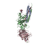
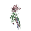
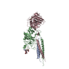
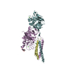
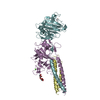
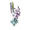
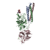
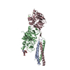
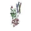
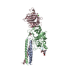
- PDB-2q9i: Crystal Structure of D-Dimer from Human Fibrin Complexed with Met... -
+ Open data

- Basic information
| Entry | Database: PDB / ID: 2q9i | |||||||||
|---|---|---|---|---|---|---|---|---|---|---|
| Title | Crystal Structure of D-Dimer from Human Fibrin Complexed with Met-His-Arg-Pro-Tyr-amide. | |||||||||
 Components Components |
| |||||||||
 Keywords Keywords |  BLOOD CLOTTING / fibrin clots / B-knobs / beta-holes / Blood coagulation / Disease mutation / Glycoprotein / Phosphoprotein / Secreted / Pyrrolidone carboxylic acid BLOOD CLOTTING / fibrin clots / B-knobs / beta-holes / Blood coagulation / Disease mutation / Glycoprotein / Phosphoprotein / Secreted / Pyrrolidone carboxylic acid | |||||||||
| Function / homology |  Function and homology information Function and homology informationplatelet maturation / blood coagulation, common pathway / induction of bacterial agglutination / fibrinogen complex / Regulation of TLR by endogenous ligand / platelet alpha granule / blood coagulation, fibrin clot formation / cellular response to leptin stimulus / cellular response to interleukin-6 / MyD88 deficiency (TLR2/4) ...platelet maturation / blood coagulation, common pathway / induction of bacterial agglutination / fibrinogen complex / Regulation of TLR by endogenous ligand / platelet alpha granule / blood coagulation, fibrin clot formation / cellular response to leptin stimulus / cellular response to interleukin-6 / MyD88 deficiency (TLR2/4) / positive regulation of heterotypic cell-cell adhesion / IRAK4 deficiency (TLR2/4) / MyD88:MAL(TIRAP) cascade initiated on plasma membrane / extracellular matrix structural constituent / plasminogen activation / p130Cas linkage to MAPK signaling for integrins / positive regulation of peptide hormone secretion / positive regulation of exocytosis / GRB2:SOS provides linkage to MAPK signaling for Integrins / protein secretion / protein polymerization / cellular response to interleukin-1 / Integrin cell surface interactions / negative regulation of endothelial cell apoptotic process / Common Pathway of Fibrin Clot Formation / positive regulation of substrate adhesion-dependent cell spreading / negative regulation of extrinsic apoptotic signaling pathway via death domain receptors / positive regulation of vasoconstriction / cell adhesion molecule binding / fibrinolysis / Integrin signaling / cell-matrix adhesion / platelet alpha granule lumen / positive regulation of protein secretion / Post-translational protein phosphorylation / Signaling by high-kinase activity BRAF mutants / MAP2K and MAPK activation / platelet aggregation / response to calcium ion / Signaling by RAF1 mutants / Signaling by moderate kinase activity BRAF mutants / Paradoxical activation of RAF signaling by kinase inactive BRAF / Signaling downstream of RAS mutants / Regulation of Insulin-like Growth Factor (IGF) transport and uptake by Insulin-like Growth Factor Binding Proteins (IGFBPs) / extracellular vesicle / Signaling by BRAF and RAF1 fusions / Platelet degranulation / cell cortex / ER-Phagosome pathway / protein-folding chaperone binding / protein-containing complex assembly / collagen-containing extracellular matrix / adaptive immune response / blood microparticle / positive regulation of ERK1 and ERK2 cascade / Amyloid fiber formation / external side of plasma membrane / endoplasmic reticulum lumen / signaling receptor binding / innate immune response / synapse / structural molecule activity / cell surface / endoplasmic reticulum / extracellular space / extracellular exosome / extracellular region / identical protein binding / metal ion binding / plasma membraneSimilarity search - Function | |||||||||
| Biological species |  Homo sapiens (human) Homo sapiens (human) | |||||||||
| Method | X-RAY DIFFRACTION / MOLECULAR REPLACEMENT / Resolution: 2.8 Å | |||||||||
 Authors Authors | Doolittle, R.F. / Pandi, L. | |||||||||
 Citation Citation | Journal: Biochemistry / Year: 2007 Title: Probing the beta-chain hole of fibrinogen with synthetic peptides that differ at their amino termini Authors: Doolittle, R.F. / Pandi, L. | |||||||||
| History |
|
- Structure visualization
| Structure viewer | Molecule: MolmilJmol/JSmol |
|---|
- Downloads & links
-Download
| PDBx/mmCIF format | 2q9i.cif.gz | 252.7 KB | Display | PDBx/mmCIF format |
|---|---|---|---|---|
| PDB format | pdb2q9i.ent.gz | 204.2 KB | Display | PDB format |
| PDBx/mmJSON format | 2q9i.json.gz | Tree view | PDBx/mmJSON format | |
| Others |  Other downloads Other downloads |
-Validation report
| Arichive directory | https://data.pdbj.org/pub/pdb/validation_reports/q9/2q9iftp://data.pdbj.org/pub/pdb/validation_reports/q9/2q9i | HTTPS FTP |
|---|
-Related structure data
| Related structure data | 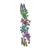 2z4eC 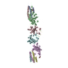 1fzfS S: Starting model for refinement C: citing same article ( |
|---|---|
| Similar structure data |


-Links
 PDBj
PDBj


- Assembly
| Deposited unit | 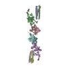
| ||||||||
|---|---|---|---|---|---|---|---|---|---|
| 1 | 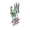
| ||||||||
| 2 | 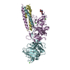
| ||||||||
| Unit cell |
|
-Components
-Protein , 3 types, 6 molecules ADBECF
| #1: Protein | Mass: 10244.963 Da / Num. of mol.: 2 / Fragment: UNP residues 130-216 / Source method: isolated from a natural source / Source: (natural) Homo sapiens (human) / References: UniProt: P02671#2: Protein | Mass: 37691.992 Da / Num. of mol.: 2 / Fragment: UNP residues 164-491 / Source method: isolated from a natural source / Source: (natural) Homo sapiens (human) / References: UniProt: P02675#3: Protein | / Fibrinogen gamma chain / isoform CRA_mMass: 36693.754 Da / Num. of mol.: 2 / Fragment: UNP residues 114-437 / Source method: isolated from a natural source / Source: (natural) Homo sapiens (human) / References: UniProt: Q53Y18, UniProt: P02679*PLUS |
|---|
-Fibrin B Knob ... , 2 types, 4 molecules STMN
| #4: Protein/peptide | Mass: 467.522 Da / Num. of mol.: 2 / Source method: obtained synthetically / Details: This sequence occurs naturally in humans. #5: Protein/peptide | Mass: 704.840 Da / Num. of mol.: 2 / Source method: obtained synthetically / Details: This sequence does not occur naturally. |
|---|
-Sugars / Non-polymers , 2 types, 10 molecules 
| #6: Polysaccharide | / Mass: 424.401 Da / Num. of mol.: 2 Source method: isolated from a genetically manipulated source #7: Chemical | ChemComp-CA /  Mass: 40.078 Da / Num. of mol.: 8 / Source method: obtained synthetically / Formula: Ca Mass: 40.078 Da / Num. of mol.: 8 / Source method: obtained synthetically / Formula: Ca |
|---|
-Experimental details
-Experiment
| Experiment | Method: X-RAY DIFFRACTION / Number of used crystals: 1 |
|---|
- Sample preparation
| Crystal | Density Matthews: 2.7 Å3/Da / Density % sol: 54.44 % |
|---|---|
| Crystal grow | Temperature: 295 K / Method: vapor diffusion, sitting drop / pH: 7.5 Details: equal volumes of (a) 9 mg/ml D-dimer, 2 mM Gly-His-Arg-Pro-amide, 0.05 M Tris, pH 7.0, 5 mM CaCl2 and (b) 10% PEG, 5 mM CaCl2, 0.05M Tris, pH 8.0, 2 mM sodium azide. Crystals were ...Details: equal volumes of (a) 9 mg/ml D-dimer, 2 mM Gly-His-Arg-Pro-amide, 0.05 M Tris, pH 7.0, 5 mM CaCl2 and (b) 10% PEG, 5 mM CaCl2, 0.05M Tris, pH 8.0, 2 mM sodium azide. Crystals were subsequently soaked in same solution containing 0.6 mM Met-His-Arg-Pro-Tyr-amide., pH 7.5, VAPOR DIFFUSION, SITTING DROP, temperature 295.0K |
-Data collection
| Diffraction | Mean temperature: 100 K |
|---|---|
| Diffraction source | Source: ROTATING ANODE / Type: RIGAKU / Wavelength: 1.54 Å |
| Detector | Type: MAR scanner 345 mm plate / Detector: IMAGE PLATE / Date: Nov 15, 2006 / Details: Confocal Max-Flux Optic |
| Radiation | Monochromator: osmic / Protocol: SINGLE WAVELENGTH / Monochromatic (M) / Laue (L): M / Scattering type: x-ray |
| Radiation wavelength | Wavelength: 1.54 Å / Relative weight: 1 |
| Reflection | Resolution: 2.8→50 Å / Num. all: 46081 / Num. obs: 44180 / % possible obs: 95.4 % / Observed criterion σ(F): 0 / Observed criterion σ(I): 0 / Redundancy: 3.5 % / Rsym value: 0.085 |
| Reflection shell | Resolution: 2.8→2.9 Å / Redundancy: 2.5 % / Rsym value: 0.572 / % possible all: 83.5 |
- Processing
| Software |
| ||||||||||||||||||||||||||||||||||||||||||||||||||||||||||||||||||||||||||||||||
|---|---|---|---|---|---|---|---|---|---|---|---|---|---|---|---|---|---|---|---|---|---|---|---|---|---|---|---|---|---|---|---|---|---|---|---|---|---|---|---|---|---|---|---|---|---|---|---|---|---|---|---|---|---|---|---|---|---|---|---|---|---|---|---|---|---|---|---|---|---|---|---|---|---|---|---|---|---|---|---|---|---|
| Refinement | Method to determine structure: MOLECULAR REPLACEMENT Starting model: 1FZF Resolution: 2.8→30 Å / Cross valid method: THROUGHOUT / σ(F): 1
| ||||||||||||||||||||||||||||||||||||||||||||||||||||||||||||||||||||||||||||||||
| Solvent computation | Bsol: 39.446 Å2 | ||||||||||||||||||||||||||||||||||||||||||||||||||||||||||||||||||||||||||||||||
| Displacement parameters | Biso mean: 40.6025 Å2
| ||||||||||||||||||||||||||||||||||||||||||||||||||||||||||||||||||||||||||||||||
| Refinement step | Cycle: LAST / Resolution: 2.8→30 Å
| ||||||||||||||||||||||||||||||||||||||||||||||||||||||||||||||||||||||||||||||||
| Refine LS restraints |
| ||||||||||||||||||||||||||||||||||||||||||||||||||||||||||||||||||||||||||||||||
| Xplor file |
|
