 Movie
Movie Controller

[English] 日本語
 Yorodumi
- PDB-1kma: NMR Structure of the Domain-I of the Kazal-type Thrombin Inhibito... -
+ Open data

- Basic information
| Entry | Database: PDB / ID: 1kma | ||||||
|---|---|---|---|---|---|---|---|
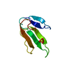
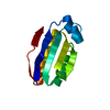
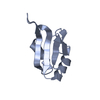
| Title | NMR Structure of the Domain-I of the Kazal-type Thrombin Inhibitor Dipetalin | ||||||
 Components Components | DIPETALIN | ||||||
 Keywords Keywords |  BLOOD CLOTTING / disulphide-rich small alpha+beta fold / Kazal-type BLOOD CLOTTING / disulphide-rich small alpha+beta fold / Kazal-type | ||||||
| Function / homology |  Function and homology information Function and homology informationnegative regulation of coagulation / serine-type endopeptidase inhibitor activity / extracellular region Similarity search - Function | ||||||
| Biological species |  Dipetalogaster maximus (insect) Dipetalogaster maximus (insect) | ||||||
| Method | SOLUTION NMR / distance geometry, simulated annealing | ||||||
 Authors Authors | Schlott, B. / Wohnert, J. / Icke, C. / Hartmann, M. / Ramachandran, R. / Guhrs, K.-H. / Glusa, E. / Flemming, J. / Gorlach, M. / Grosse, F. / Ohlenschlager, O. | ||||||
 Citation Citation | Journal: J.Mol.Biol. / Year: 2002 Title: Interaction of Kazal-type inhibitor domains with serine proteinases: biochemical and structural studies. Authors: Schlott, B. / Wohnert, J. / Icke, C. / Hartmann, M. / Ramachandran, R. / Guhrs, K.H. / Glusa, E. / Flemming, J. / Gorlach, M. / Grosse, F. / Ohlenschlager, O. | ||||||
| History |
|
- Structure visualization
| Structure viewer | Molecule: MolmilJmol/JSmol |
|---|
- Downloads & links
-Download
| PDBx/mmCIF format | 1kma.cif.gz | 306.2 KB | Display | PDBx/mmCIF format |
|---|---|---|---|---|
| PDB format | pdb1kma.ent.gz | 265 KB | Display | PDB format |
| PDBx/mmJSON format | 1kma.json.gz | Tree view | PDBx/mmJSON format | |
| Others |  Other downloads Other downloads |
-Validation report
| Arichive directory | https://data.pdbj.org/pub/pdb/validation_reports/km/1kmaftp://data.pdbj.org/pub/pdb/validation_reports/km/1kma | HTTPS FTP |
|---|
-Related structure data
| Similar structure data |
|---|


-Links
 PDBj
PDBj
- Assembly
| Deposited unit | 
| |||||||||
|---|---|---|---|---|---|---|---|---|---|---|
| 1 |
| |||||||||
| NMR ensembles |
|
-Components
| #1: Protein | Mass: 6090.670 Da / Num. of mol.: 1 / Fragment: N-TERMINAL DOMAIN-I Source method: isolated from a genetically manipulated source Source: (gene. exp.) Dipetalogaster maximus (insect) / Plasmid: pMEX6 / Production host:  Escherichia coli (E. coli) / Strain (production host): TG1 / References: UniProt: O96790 Escherichia coli (E. coli) / Strain (production host): TG1 / References: UniProt: O96790 |
|---|
-Experimental details
-Experiment
| Experiment | Method: SOLUTION NMR | ||||||||||||||||
|---|---|---|---|---|---|---|---|---|---|---|---|---|---|---|---|---|---|
| NMR experiment |
| ||||||||||||||||
| NMR details | Text: The structure was determined using triple-resonance NMR spectroscopy. |
- Sample preparation
| Details |
| |||||||||
|---|---|---|---|---|---|---|---|---|---|---|
| Sample conditions | pH: 6.68 / Pressure: ambient / Temperature: 288 K | |||||||||
| Crystal grow | *PLUS Method: other / Details: NMR |
-NMR measurement
| Radiation | Protocol: SINGLE WAVELENGTH / Monochromatic (M) / Laue (L): M | |||||||||||||||
|---|---|---|---|---|---|---|---|---|---|---|---|---|---|---|---|---|
| Radiation wavelength | Relative weight: 1 | |||||||||||||||
| NMR spectrometer |
|
- Processing
| NMR software |
| ||||||||||||
|---|---|---|---|---|---|---|---|---|---|---|---|---|---|
| Refinement | Method: distance geometry, simulated annealing / Software ordinal: 1 Details: The structures are based on a total of 1122 distance constraints and 239 dihedral angle restraints. | ||||||||||||
| NMR representative | Selection criteria: closest to the average | ||||||||||||
| NMR ensemble | Conformer selection criteria: target function / Conformers calculated total number: 100 / Conformers submitted total number: 20 |
