 Movie
Movie Controller

[English] 日本語
 Yorodumi
- PDB-3hi9: The x-ray crystal structure of the first RNA recognition motif (R... -
+ Open data

- Basic information
| Entry | Database: PDB / ID: 3hi9 | ||||||
|---|---|---|---|---|---|---|---|
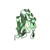
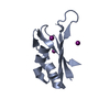
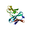
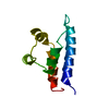
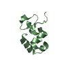
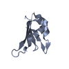
| Title | The x-ray crystal structure of the first RNA recognition motif (RRM1) of the AU-rich element (ARE) binding protein HuR at 2.0 angstrom resolution | ||||||
 Components Components | ELAV-like protein 1 | ||||||
 Keywords Keywords | TRANSCRIPTION / beta1-alpha1-beta2-beta3-alpha2-beta4 topology / Methylation / Phosphoprotein / RNA-binding | ||||||
| Function / homology |  Function and homology information Function and homology informationHuR (ELAVL1) binds and stabilizes mRNA / negative regulation of miRNA-mediated gene silencing / post-transcriptional gene silencing / regulation of stem cell population maintenance / mRNA 3'-UTR AU-rich region binding / mRNA stabilization / lncRNA binding / miRNA binding / 3'-UTR-mediated mRNA stabilization / mRNA destabilization ...HuR (ELAVL1) binds and stabilizes mRNA / negative regulation of miRNA-mediated gene silencing / post-transcriptional gene silencing / regulation of stem cell population maintenance / mRNA 3'-UTR AU-rich region binding / mRNA stabilization / lncRNA binding / miRNA binding / 3'-UTR-mediated mRNA stabilization / mRNA destabilization / sarcoplasm / positive regulation of superoxide anion generation / mRNA 3'-UTR binding / positive regulation of translation / P-body / protein homooligomerization / cytoplasmic stress granule / protein import into nucleus / double-stranded RNA binding / cytoplasmic vesicle / postsynapse / ribonucleoprotein complex / mRNA binding / glutamatergic synapse / protein kinase binding / endoplasmic reticulum / protein homodimerization activity / RNA binding / nucleoplasm / membrane / nucleus / cytosol / cytoplasmSimilarity search - Function | ||||||
| Biological species |  Homo sapiens (human) Homo sapiens (human) | ||||||
| Method | X-RAY DIFFRACTION / SYNCHROTRON / MOLECULAR REPLACEMENT / molecular replacement / Resolution: 2 Å | ||||||
 Authors Authors | Benoit, R.M. / Kallen, J. | ||||||
 Citation Citation | Journal: J.Mol.Biol. / Year: 2010 Title: The X-ray Crystal Structure of the First RNA Recognition Motif and Site-Directed Mutagenesis Suggest a Possible HuR Redox Sensing Mechanism. Authors: Benoit, R.M. / Meisner, N.C. / Kallen, J. / Graff, P. / Hemmig, R. / Cebe, R. / Ostermeier, C. / Widmer, H. / Auer, M. | ||||||
| History |
|
- Structure visualization
| Structure viewer | Molecule: MolmilJmol/JSmol |
|---|
- Downloads & links
-Download
| PDBx/mmCIF format | 3hi9.cif.gz | 76.9 KB | Display | PDBx/mmCIF format |
|---|---|---|---|---|
| PDB format | pdb3hi9.ent.gz | 58.8 KB | Display | PDB format |
| PDBx/mmJSON format | 3hi9.json.gz | Tree view | PDBx/mmJSON format | |
| Others |  Other downloads Other downloads |
-Validation report
| Arichive directory | https://data.pdbj.org/pub/pdb/validation_reports/hi/3hi9ftp://data.pdbj.org/pub/pdb/validation_reports/hi/3hi9 | HTTPS FTP |
|---|
-Related structure data
| Related structure data | 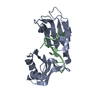 1fxlS S: Starting model for refinement |
|---|---|
| Similar structure data |

-Links
 PDBj
PDBj
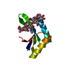
- Assembly
| Deposited unit | 
| ||||||||
|---|---|---|---|---|---|---|---|---|---|
| 1 | 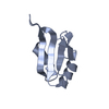
| ||||||||
| 2 | 
| ||||||||
| 3 | 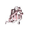
| ||||||||
| 4 | 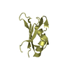
| ||||||||
| Unit cell |
|
-Components
| #1: Protein | / Hu-antigen R / HuR Mass: 9252.491 Da / Num. of mol.: 4 / Fragment: RRM1 domain: UNP residues 18-99 Source method: isolated from a genetically manipulated source Source: (gene. exp.) Homo sapiens (human) / Gene: ELAVL1, HUR / Plasmid: pET28a / Production host:  Escherichia coli (E. coli) / Strain (production host): Rosetta2(DE3) / References: UniProt: Q15717 Escherichia coli (E. coli) / Strain (production host): Rosetta2(DE3) / References: UniProt: Q15717#2: Water | ChemComp-HOH / | Water Mass: 18.015 Da / Num. of mol.: 197 / Source method: isolated from a natural source / Formula: H2O Mass: 18.015 Da / Num. of mol.: 197 / Source method: isolated from a natural source / Formula: H2O |
|---|
-Experimental details
-Experiment
| Experiment | Method: X-RAY DIFFRACTION / Number of used crystals: 1 |
|---|
- Sample preparation
| Crystal | Density Matthews: 3.22 Å3/Da / Density % sol: 61.75 % |
|---|---|
| Crystal grow | Temperature: 293 K / Method: vapor diffusion, hanging drop / pH: 5.6 Details: 0.2 M Di-ammonium citrate, 20% w/v PEG 3350, 5% Glycerol, pH 5.6, VAPOR DIFFUSION, HANGING DROP, temperature 293K |
-Data collection
| Diffraction | Mean temperature: 100 K |
|---|---|
| Diffraction source | Source: SYNCHROTRON / Site: SLS  / Beamline: X10SA / Wavelength: 1.00778 Å / Beamline: X10SA / Wavelength: 1.00778 Å |
| Detector | Type: MARMOSAIC 225 mm CCD / Detector: CCD / Date: Jun 22, 2006 |
| Radiation | Protocol: SINGLE WAVELENGTH / Monochromatic (M) / Laue (L): M / Scattering type: x-ray |
| Radiation wavelength | Wavelength: 1.00778 Å / Relative weight: 1 |
| Reflection | Resolution: 2→30 Å / Num. obs: 32767 / % possible obs: 99.3 % / Observed criterion σ(I): -3 / Redundancy: 3 % / Biso Wilson estimate: 33.895 Å2 / Rmerge(I) obs: 0.096 / Net I/σ(I): 7.9 |
| Reflection shell | Resolution: 2→2.08 Å / Redundancy: 3 % / Rmerge(I) obs: 0.484 / Mean I/σ(I) obs: 3.2 / Num. measured obs: 11167 / Num. unique all: 3709 / Num. unique obs: 3709 / % possible all: 99.5 |
-Phasing
| Phasing | Method: molecular replacement |
|---|
- Processing
| Software |
| |||||||||||||||||||||||||||||||||||||||||||||||||||||||||||||||||
|---|---|---|---|---|---|---|---|---|---|---|---|---|---|---|---|---|---|---|---|---|---|---|---|---|---|---|---|---|---|---|---|---|---|---|---|---|---|---|---|---|---|---|---|---|---|---|---|---|---|---|---|---|---|---|---|---|---|---|---|---|---|---|---|---|---|---|
| Refinement | Method to determine structure: MOLECULAR REPLACEMENT Starting model: PDB entry 1FXL Resolution: 2→29.76 Å / Cor.coef. Fo:Fc: 0.953 / Cor.coef. Fo:Fc free: 0.937 / WRfactor Rfree: 0.237 / WRfactor Rwork: 0.2 / Occupancy max: 1 / Occupancy min: 0.5 / FOM work R set: 0.847 / SU B: 3.764 / SU ML: 0.104 / SU R Cruickshank DPI: 0.147 / SU Rfree: 0.139 / Cross valid method: THROUGHOUT / σ(F): 0 / ESU R: 0.147 / ESU R Free: 0.139 / Stereochemistry target values: MAXIMUM LIKELIHOOD Details: 1. HYDROGENS HAVE BEEN ADDED IN THE RIDING POSITIONS. 2. U VALUES: REFINED INDIVIDUALLY.
| |||||||||||||||||||||||||||||||||||||||||||||||||||||||||||||||||
| Solvent computation | Ion probe radii: 0.8 Å / Shrinkage radii: 0.8 Å / VDW probe radii: 1.4 Å / Solvent model: MASK | |||||||||||||||||||||||||||||||||||||||||||||||||||||||||||||||||
| Displacement parameters | Biso max: 67.53 Å2 / Biso mean: 32.295 Å2 / Biso min: 16.13 Å2
| |||||||||||||||||||||||||||||||||||||||||||||||||||||||||||||||||
| Refinement step | Cycle: LAST / Resolution: 2→29.76 Å
| |||||||||||||||||||||||||||||||||||||||||||||||||||||||||||||||||
| Refine LS restraints |
| |||||||||||||||||||||||||||||||||||||||||||||||||||||||||||||||||
| LS refinement shell | Resolution: 2→2.052 Å / Total num. of bins used: 20
|
