 Movie
Movie Controller

[English] 日本語
 Yorodumi
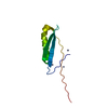
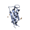
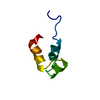
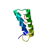
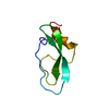
- PDB-1k50: A V49A Mutation Induces 3D Domain Swapping in the B1 Domain of Pr... -
+ Open data

- Basic information
| Entry | Database: PDB / ID: 1k50 | ||||||
|---|---|---|---|---|---|---|---|
| Title | A V49A Mutation Induces 3D Domain Swapping in the B1 Domain of Protein L from Peptostreptococcus magnus | ||||||
 Components Components | Protein L | ||||||
 Keywords Keywords | PROTEIN BINDING / Protein L B1 domain / strained beta-hairpin turn / positive phi angles / domain swapping / amyloid formation | ||||||
| Function / homology |  Function and homology information Function and homology information | ||||||
| Biological species |  Finegoldia magna (bacteria) Finegoldia magna (bacteria) | ||||||
| Method | X-RAY DIFFRACTION / MOLECULAR REPLACEMENT / Resolution: 1.8 Å | ||||||
 Authors Authors | O'Neill, J.W. / Kim, D.E. / Johnsen, K. / Baker, D. / Zhang, K.Y.J. | ||||||
 Citation Citation | Journal: Structure / Year: 2001 Title: Single-site mutations induce 3D domain swapping in the B1 domain of protein L from Peptostreptococcus magnus. Authors: O'Neill, J.W. / Kim, D.E. / Johnsen, K. / Baker, D. / Zhang, K.Y. | ||||||
| History |
|
- Structure visualization
| Structure viewer | Molecule: MolmilJmol/JSmol |
|---|
- Downloads & links
-Download
| PDBx/mmCIF format | 1k50.cif.gz | 61.9 KB | Display | PDBx/mmCIF format |
|---|---|---|---|---|
| PDB format | pdb1k50.ent.gz | 46.9 KB | Display | PDB format |
| PDBx/mmJSON format | 1k50.json.gz | Tree view | PDBx/mmJSON format | |
| Others |  Other downloads Other downloads |
-Validation report
| Arichive directory | https://data.pdbj.org/pub/pdb/validation_reports/k5/1k50ftp://data.pdbj.org/pub/pdb/validation_reports/k5/1k50 | HTTPS FTP |
|---|
-Related structure data
| Related structure data | 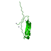 1k51C  1k52C  1k53C  1hz6S S: Starting model for refinement C: citing same article ( |
|---|---|
| Similar structure data |


-Links
 PDBj
PDBj- Assembly
| Deposited unit | 
| ||||||||
|---|---|---|---|---|---|---|---|---|---|
| 1 | 
| ||||||||
| 2 | 
| ||||||||
| 3 | 
| ||||||||
| 4 |
| ||||||||
| Unit cell |
| ||||||||
| Details | 2 monomers(Chain A,C)and 1 domain swapped dimer (Chain B,D)in asymmetric unit |
-Components
| #1: Protein | Mass: 6862.503 Da / Num. of mol.: 4 / Fragment: B1 Domain (Residues 111-173) / Mutation: V49A, Y47W Source method: isolated from a genetically manipulated source Source: (gene. exp.) Finegoldia magna (bacteria) / Strain: ATCC 29328 / Gene: Protein L, B1 domain / Plasmid: pET3a / Species (production host): Escherichia coli / Production host: Escherichia coli BL21(DE3) (bacteria) / Strain (production host): BL21(DE3) / References: UniProt: Q51912#2: Water | ChemComp-HOH / | Water Mass: 18.015 Da / Num. of mol.: 182 / Source method: isolated from a natural source / Formula: H2O Mass: 18.015 Da / Num. of mol.: 182 / Source method: isolated from a natural source / Formula: H2O |
|---|
-Experimental details
-Experiment
| Experiment | Method: X-RAY DIFFRACTION / Number of used crystals: 1 |
|---|
- Sample preparation
| Crystal | Density Matthews: 2.97 Å3/Da / Density % sol: 58.59 % | ||||||||||||||||||||||||||||||
|---|---|---|---|---|---|---|---|---|---|---|---|---|---|---|---|---|---|---|---|---|---|---|---|---|---|---|---|---|---|---|---|
| Crystal grow | Temperature: 277 K / Method: vapor diffusion, hanging drop / pH: 4.5 Details: 18% PEG3350, 0.2M (NH4)2SO4, 100mM Citrate, pH 4.5, VAPOR DIFFUSION, HANGING DROP, temperature 277K | ||||||||||||||||||||||||||||||
| Crystal grow | *PLUS pH: 7 | ||||||||||||||||||||||||||||||
| Components of the solutions | *PLUS
|
-Data collection
| Diffraction | Mean temperature: 100 K |
|---|---|
| Diffraction source | Source: ROTATING ANODE / Type: RIGAKU / Wavelength: 1.5418 Å |
| Detector | Type: RIGAKU RAXIS IV / Detector: IMAGE PLATE / Date: Jun 25, 1999 / Details: mirrors |
| Radiation | Monochromator: Yale mirrors / Protocol: SINGLE WAVELENGTH / Monochromatic (M) / Laue (L): M / Scattering type: x-ray |
| Radiation wavelength | Wavelength: 1.5418 Å / Relative weight: 1 |
| Reflection | Resolution: 1.8→25 Å / Num. all: 28770 / Num. obs: 28396 / % possible obs: 95.9 % / Observed criterion σ(F): 0 / Observed criterion σ(I): -3 / Redundancy: 4.7 % / Biso Wilson estimate: 21.8 Å2 / Rmerge(I) obs: 0.047 / Rsym value: 0.04 / Net I/σ(I): 30.9 |
| Reflection shell | Resolution: 1.8→1.86 Å / Rmerge(I) obs: 0.435 / Mean I/σ(I) obs: 2.3 / Rsym value: 0.418 / % possible all: 93.3 |
| Reflection | *PLUS Lowest resolution: 25 Å / Num. obs: 29286 / % possible obs: 98.7 % / Num. measured all: 134022 / Rmerge(I) obs: 0.047 |
| Reflection shell | *PLUS Rmerge(I) obs: 0.435 |
- Processing
| Software |
| ||||||||||||||||||||||||||||||||||||
|---|---|---|---|---|---|---|---|---|---|---|---|---|---|---|---|---|---|---|---|---|---|---|---|---|---|---|---|---|---|---|---|---|---|---|---|---|---|
| Refinement | Method to determine structure: MOLECULAR REPLACEMENT Starting model: PDB ENTRY 1hz6 Resolution: 1.8→24.14 Å / Rfactor Rfree error: 0.005 / Data cutoff high absF: 441418.03 / Data cutoff low absF: 0 / Isotropic thermal model: RESTRAINED / Cross valid method: THROUGHOUT / σ(F): 0 / Stereochemistry target values: maximum likelyhood
| ||||||||||||||||||||||||||||||||||||
| Solvent computation | Solvent model: FLAT MODEL / Bsol: 52.6397 Å2 / ksol: 0.378529 e/Å3 | ||||||||||||||||||||||||||||||||||||
| Displacement parameters | Biso mean: 31.4 Å2
| ||||||||||||||||||||||||||||||||||||
| Refine analyze |
| ||||||||||||||||||||||||||||||||||||
| Refinement step | Cycle: LAST / Resolution: 1.8→24.14 Å
| ||||||||||||||||||||||||||||||||||||
| Refine LS restraints |
| ||||||||||||||||||||||||||||||||||||
| LS refinement shell | Resolution: 1.8→1.91 Å / Rfactor Rfree error: 0.015 / Total num. of bins used: 6
| ||||||||||||||||||||||||||||||||||||
| Xplor file |
| ||||||||||||||||||||||||||||||||||||
| Refinement | *PLUS % reflection Rfree: 9.5 % / Rfactor obs: 0.211 / Rfactor Rfree: 0.239 / Rfactor Rwork: 0.211 | ||||||||||||||||||||||||||||||||||||
| Solvent computation | *PLUS | ||||||||||||||||||||||||||||||||||||
| Displacement parameters | *PLUS | ||||||||||||||||||||||||||||||||||||
| Refine LS restraints | *PLUS
| ||||||||||||||||||||||||||||||||||||
| LS refinement shell | *PLUS Rfactor Rfree: 0.303 / Rfactor Rwork: 0.297 |
