 Movie
Movie Controller

[English] 日本語
 Yorodumi
- PDB-1jr1: Crystal structure of Inosine Monophosphate Dehydrogenase in compl... -
+ Open data

- Basic information
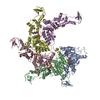
| Entry | Database: PDB / ID: 1jr1 | ||||||
|---|---|---|---|---|---|---|---|
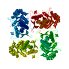
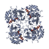
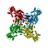
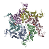
| Title | Crystal structure of Inosine Monophosphate Dehydrogenase in complex with Mycophenolic Acid | ||||||
 Components Components | Inosine-5'-Monophosphate Dehydrogenase 2 | ||||||
 Keywords Keywords |  OXIDOREDUCTASE / DEHYDROGENASE / IMPD / IMPDH / GUANINE NUCLEOTIDE SYNTHESIS / MYCOPHENOLIC ACID / MPA OXIDOREDUCTASE / DEHYDROGENASE / IMPD / IMPDH / GUANINE NUCLEOTIDE SYNTHESIS / MYCOPHENOLIC ACID / MPA | ||||||
| Function / homology |  Function and homology information Function and homology informationlymphocyte proliferation / IMP dehydrogenase activity / IMP dehydrogenase / GMP biosynthetic process / cellular response to interleukin-4 / circadian rhythm / nucleotide binding / metal ion binding / nucleus / cytosol / cytoplasmSimilarity search - Function | ||||||
| Biological species |   Cricetulus griseus (Chinese hamster) Cricetulus griseus (Chinese hamster) | ||||||
| Method | X-RAY DIFFRACTION / MIR / Resolution: 2.6 Å | ||||||
 Authors Authors | Sintchak, M.D. / Fleming, M.A. / Futer, O. / Raybuck, S.A. / Chambers, S.P. / Caron, P.R. / Murcko, M.A. / Wilson, K.P. | ||||||
 Citation Citation | Journal: Cell(Cambridge,Mass.) / Year: 1996 Title: Structure and mechanism of inosine monophosphate dehydrogenase in complex with the immunosuppressant mycophenolic acid. Authors: Sintchak, M.D. / Fleming, M.A. / Futer, O. / Raybuck, S.A. / Chambers, S.P. / Caron, P.R. / Murcko, M.A. / Wilson, K.P. | ||||||
| History |
|
- Structure visualization
| Structure viewer | Molecule: MolmilJmol/JSmol |
|---|
- Downloads & links
-Download
| PDBx/mmCIF format | 1jr1.cif.gz | 165.9 KB | Display | PDBx/mmCIF format |
|---|---|---|---|---|
| PDB format | pdb1jr1.ent.gz | 135.8 KB | Display | PDB format |
| PDBx/mmJSON format | 1jr1.json.gz | Tree view | PDBx/mmJSON format | |
| Others |  Other downloads Other downloads |
-Validation report
| Arichive directory | https://data.pdbj.org/pub/pdb/validation_reports/jr/1jr1ftp://data.pdbj.org/pub/pdb/validation_reports/jr/1jr1 | HTTPS FTP |
|---|
-Related structure data
| Related structure data | |
|---|---|
| Similar structure data |


-Links
 PDBj
PDBj
- Assembly
| Deposited unit | 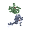
| ||||||||
|---|---|---|---|---|---|---|---|---|---|
| 1 | 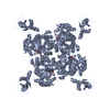
| ||||||||
| 2 | 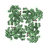
| ||||||||
| Unit cell |
| ||||||||
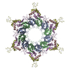
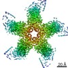
| Details | the biological assembly is a tetramer generated from chain A in the asymmetric unit by the operations: -x,-y,z; -y,x,z; y,-x,z |
-Components
| #1: Protein | Mass: 55961.082 Da / Num. of mol.: 2 Source method: isolated from a genetically manipulated source Source: (gene. exp.) Cricetulus griseus (Chinese hamster) / Production host:  Escherichia coli (E. coli) / References: UniProt: P12269, IMP dehydrogenase Escherichia coli (E. coli) / References: UniProt: P12269, IMP dehydrogenase#2: Chemical |   Mass: 39.098 Da / Num. of mol.: 2 / Source method: obtained synthetically / Formula: K Mass: 39.098 Da / Num. of mol.: 2 / Source method: obtained synthetically / Formula: K#3: Chemical | Inosinic acid  Mass: 348.206 Da / Num. of mol.: 2 / Source method: obtained synthetically / Formula: C10H13N4O8P Mass: 348.206 Da / Num. of mol.: 2 / Source method: obtained synthetically / Formula: C10H13N4O8P#4: Chemical | Mycophenolic acid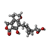  Mass: 320.337 Da / Num. of mol.: 2 / Source method: obtained synthetically / Formula: C17H20O6 / Comment: medication, immunosuppressant*YM Mass: 320.337 Da / Num. of mol.: 2 / Source method: obtained synthetically / Formula: C17H20O6 / Comment: medication, immunosuppressant*YM#5: Water | ChemComp-HOH / | Water Mass: 18.015 Da / Num. of mol.: 202 / Source method: isolated from a natural source / Formula: H2O Mass: 18.015 Da / Num. of mol.: 202 / Source method: isolated from a natural source / Formula: H2O |
|---|
-Experimental details
-Experiment
| Experiment | Method: X-RAY DIFFRACTION / Number of used crystals: 1 |
|---|
- Sample preparation
| Crystal | Density Matthews: 3.03 Å3/Da / Density % sol: 59.43 % | ||||||||||||||||||||||||||||||||||||||||||||||||||||||||||||||||||||||||||||||
|---|---|---|---|---|---|---|---|---|---|---|---|---|---|---|---|---|---|---|---|---|---|---|---|---|---|---|---|---|---|---|---|---|---|---|---|---|---|---|---|---|---|---|---|---|---|---|---|---|---|---|---|---|---|---|---|---|---|---|---|---|---|---|---|---|---|---|---|---|---|---|---|---|---|---|---|---|---|---|---|
| Crystal grow | Temperature: 298 K / Method: vapor diffusion, hanging drop / pH: 5.8 Details: PEG 6000, LiCl, MES, 1-methyl-2-pyrrolidinone, 2-mercaptoethanol, pH 5.8, VAPOR DIFFUSION, HANGING DROP, temperature 298K | ||||||||||||||||||||||||||||||||||||||||||||||||||||||||||||||||||||||||||||||
| Crystal grow | *PLUS Temperature: 22 ℃ / pH: 8 / Method: vapor diffusion | ||||||||||||||||||||||||||||||||||||||||||||||||||||||||||||||||||||||||||||||
| Components of the solutions | *PLUS
|
-Data collection
| Diffraction | Mean temperature: 100 K |
|---|---|
| Diffraction source | Source: ROTATING ANODE / Type: RIGAKU RU200 / Wavelength: 1.5418 Å |
| Detector | Type: RIGAKU RAXIS IIC / Detector: IMAGE PLATE / Date: Oct 1, 1995 |
| Radiation | Monochromator: Yale/MSC mirrors / Protocol: SINGLE WAVELENGTH / Monochromatic (M) / Laue (L): M / Scattering type: x-ray |
| Radiation wavelength | Wavelength: 1.5418 Å / Relative weight: 1 |
| Reflection | Resolution: 2.6→30 Å / Num. all: 40314 / Num. obs: 37492 / % possible obs: 93 % / Observed criterion σ(F): 1 / Observed criterion σ(I): 1 / Redundancy: 3.3 % / Rsym value: 5.2 |
| Reflection shell | Resolution: 2.6→2.71 Å / % possible all: 80 |
| Reflection | *PLUS % possible obs: 93 % / Num. measured all: 123246 / Rmerge(I) obs: 0.052 |
- Processing
| Software |
| |||||||||||||||||||||||||
|---|---|---|---|---|---|---|---|---|---|---|---|---|---|---|---|---|---|---|---|---|---|---|---|---|---|---|
| Refinement | Method to determine structure: MIR / Resolution: 2.6→8 Å / σ(F): 1 / Stereochemistry target values: Engh & Huber
| |||||||||||||||||||||||||
| Refinement step | Cycle: LAST / Resolution: 2.6→8 Å
| |||||||||||||||||||||||||
| Refine LS restraints |
| |||||||||||||||||||||||||
| LS refinement shell | Resolution: 2.6→2.71 Å
| |||||||||||||||||||||||||
| Software | *PLUS Name: X-PLOR / Version: 3.851 / Classification: refinement | |||||||||||||||||||||||||
| Refinement | *PLUS Highest resolution: 2.6 Å / Lowest resolution: 8 Å / σ(F): 1 / % reflection Rfree: 10 % / Rfactor obs: 0.217 | |||||||||||||||||||||||||
| Solvent computation | *PLUS | |||||||||||||||||||||||||
| Displacement parameters | *PLUS | |||||||||||||||||||||||||
| Refine LS restraints | *PLUS
| |||||||||||||||||||||||||
| LS refinement shell | *PLUS Rfactor Rfree: 0.317 / % reflection Rfree: 11 % / Rfactor Rwork: 0.276 |
