 Movie
Movie Controller


+ Open data

- Basic information
| Entry | Database: PDB / ID: 1jek | ||||||
|---|---|---|---|---|---|---|---|
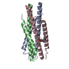
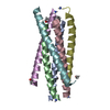
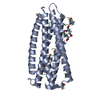
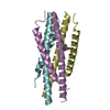
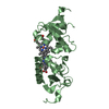
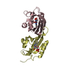
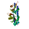
| Title | Visna TM CORE STRUCTURE | ||||||
 Components Components | (ENV POLYPROTEIN Env (gene)) x 2 Env (gene)) x 2 | ||||||
 Keywords Keywords | VIRAL PROTEIN / ENVELOPE GLYCOPROTEIN / RETROVIRUS / HIV / SIV / GP41 | ||||||
| Function / homology |  Function and homology information Function and homology informationmembrane => GO:0016020 / symbiont entry into host cell / viral envelope / virion attachment to host cell / host cell plasma membrane / virion membraneSimilarity search - Function | ||||||
| Method | X-RAY DIFFRACTION / SYNCHROTRON / MOLECULAR REPLACEMENT / Resolution: 1.5 Å | ||||||
 Authors Authors | Malashkevich, V.N. / Singh, M. / Kim, P.S. | ||||||
 Citation Citation | Journal: Proc.Natl.Acad.Sci.USA / Year: 2001 Title: The trimer-of-hairpins motif in membrane fusion: Visna virus. Authors: Malashkevich, V.N. / Singh, M. / Kim, P.S. #1: Journal: Proc.Natl.Acad.Sci.USA / Year: 1998Title: Crystal Structure of the Simian Immunodeficiency Virus (Siv) Gp41 Core: Conserved Helical Interactions Underlie the Broad Inhibitory Activity of Gp41 Peptides Authors: Malashkevich, V.N. / Chan, D.C. / Chutkowski, C.T. / Kim, P.S. #2: Journal: Cell(Cambridge,Mass.) / Year: 1997Title: Core Structure of Gp41 from the HIV Envelope Glycoprotein Authors: Chan, D.C. / Fass, D. / Berger, J.M. / Kim, P.S. | ||||||
| History |
|
- Structure visualization
| Structure viewer | Molecule: MolmilJmol/JSmol |
|---|
- Downloads & links
-Download
| PDBx/mmCIF format | 1jek.cif.gz | 30.6 KB | Display | PDBx/mmCIF format |
|---|---|---|---|---|
| PDB format | pdb1jek.ent.gz | 19.9 KB | Display | PDB format |
| PDBx/mmJSON format | 1jek.json.gz | Tree view | PDBx/mmJSON format | |
| Others |  Other downloads Other downloads |
-Validation report
| Arichive directory | https://data.pdbj.org/pub/pdb/validation_reports/je/1jekftp://data.pdbj.org/pub/pdb/validation_reports/je/1jek | HTTPS FTP |
|---|
-Related structure data
| Related structure data |  2sivS S: Starting model for refinement |
|---|---|
| Similar structure data |

-Links
 PDBj
PDBj
- Assembly
| Deposited unit | 
| ||||||||||||||||||
|---|---|---|---|---|---|---|---|---|---|---|---|---|---|---|---|---|---|---|---|
| 1 | 
| ||||||||||||||||||
| Unit cell |
| ||||||||||||||||||
| Components on special symmetry positions |
| ||||||||||||||||||
| Details | Trimer is formed around the crystallographic 3-fold axis |
-Components
| #1: Protein/peptide | Env (gene) / COAT POLYPROTEIN Mass: 4336.971 Da / Num. of mol.: 1 / Source method: obtained synthetically Details: This peptide was chemically synthesized. The sequence of the peptide is naturally found in Visna virus. References: UniProt: P35954 |
|---|---|
| #2: Protein/peptide | Env (gene) / COAT POLYPROTEIN Mass: 4126.549 Da / Num. of mol.: 1 / Source method: obtained synthetically Details: This peptide was chemically synthesized. The sequence of the peptide is naturally found in Visna virus. References: UniProt: P35954 |
| #3: Water | ChemComp-HOH / Water Mass: 18.015 Da / Num. of mol.: 142 / Source method: isolated from a natural source / Formula: H2O Mass: 18.015 Da / Num. of mol.: 142 / Source method: isolated from a natural source / Formula: H2O |
-Experimental details
-Experiment
| Experiment | Method: X-RAY DIFFRACTION / Number of used crystals: 1 |
|---|
- Sample preparation
| Crystal | Density Matthews: 2.91 Å3/Da / Density % sol: 57.67 % / Description: POLYSERINE MODEL | ||||||||||||||||||||
|---|---|---|---|---|---|---|---|---|---|---|---|---|---|---|---|---|---|---|---|---|---|
| Crystal grow | Temperature: 298 K / Method: vapor diffusion, hanging drop / pH: 8.5 Details: 25% tert-butanol, 0.1M TRIS-HCL, pH 8.5, VAPOR DIFFUSION, HANGING DROP, temperature 298K | ||||||||||||||||||||
| Crystal grow | *PLUS | ||||||||||||||||||||
| Components of the solutions | *PLUS
|
-Data collection
| Diffraction | Mean temperature: 100 K |
|---|---|
| Diffraction source | Source: SYNCHROTRON / Site: NSLS  / Beamline: X4A / Wavelength: 1.28 / Beamline: X4A / Wavelength: 1.28 |
| Detector | Type: RIGAKU RAXIS IV / Detector: IMAGE PLATE / Date: Dec 1, 1998 |
| Radiation | Protocol: SINGLE WAVELENGTH / Monochromatic (M) / Laue (L): M / Scattering type: x-ray |
| Radiation wavelength | Wavelength: 1.28 Å / Relative weight: 1 |
| Reflection | Resolution: 1.5→25 Å / Num. obs: 16300 / % possible obs: 90.1 % / Observed criterion σ(I): 0 / Redundancy: 7.5 % / Biso Wilson estimate: 22.9 Å2 / Rmerge(I) obs: 0.066 / Rsym value: 0.066 / Net I/σ(I): 13.9 |
| Reflection shell | Resolution: 1.5→1.52 Å / Redundancy: 1.5 % / Rmerge(I) obs: 0.138 / Mean I/σ(I) obs: 2.5 / Rsym value: 0.138 / % possible all: 64.7 |
| Reflection | *PLUS Num. measured all: 52353 |
| Reflection shell | *PLUS Highest resolution: 1.5 Å / % possible obs: 64.7 % / Rmerge(I) obs: 0.259 |
- Processing
| Software |
| ||||||||||||||||||||||||||||||||||||||||||||||||||||||||||||
|---|---|---|---|---|---|---|---|---|---|---|---|---|---|---|---|---|---|---|---|---|---|---|---|---|---|---|---|---|---|---|---|---|---|---|---|---|---|---|---|---|---|---|---|---|---|---|---|---|---|---|---|---|---|---|---|---|---|---|---|---|---|
| Refinement | Method to determine structure: MOLECULAR REPLACEMENT Starting model: PDB ENTRY 2SIV Resolution: 1.5→9.96 Å / Rfactor Rfree error: 0.007 / Data cutoff high absF: 914104.1 / Data cutoff low absF: 0 / Isotropic thermal model: RESTRAINED / Cross valid method: THROUGHOUT / σ(F): 0
| ||||||||||||||||||||||||||||||||||||||||||||||||||||||||||||
| Solvent computation | Solvent model: FLAT MODEL / Bsol: 94.78 Å2 / ksol: 0.451 e/Å3 | ||||||||||||||||||||||||||||||||||||||||||||||||||||||||||||
| Displacement parameters | Biso mean: 31.1 Å2
| ||||||||||||||||||||||||||||||||||||||||||||||||||||||||||||
| Refine analyze |
| ||||||||||||||||||||||||||||||||||||||||||||||||||||||||||||
| Refinement step | Cycle: LAST / Resolution: 1.5→9.96 Å
| ||||||||||||||||||||||||||||||||||||||||||||||||||||||||||||
| Refine LS restraints |
| ||||||||||||||||||||||||||||||||||||||||||||||||||||||||||||
| LS refinement shell | Resolution: 1.5→1.59 Å / Rfactor Rfree error: 0.026 / Total num. of bins used: 6
| ||||||||||||||||||||||||||||||||||||||||||||||||||||||||||||
| Xplor file |
| ||||||||||||||||||||||||||||||||||||||||||||||||||||||||||||
| Software | *PLUS Name: CNS / Version: 1 / Classification: refinement | ||||||||||||||||||||||||||||||||||||||||||||||||||||||||||||
| Refinement | *PLUS σ(F): 0 / % reflection Rfree: 10.1 % | ||||||||||||||||||||||||||||||||||||||||||||||||||||||||||||
| Solvent computation | *PLUS | ||||||||||||||||||||||||||||||||||||||||||||||||||||||||||||
| Displacement parameters | *PLUS Biso mean: 31.1 Å2 | ||||||||||||||||||||||||||||||||||||||||||||||||||||||||||||
| Refine LS restraints | *PLUS
| ||||||||||||||||||||||||||||||||||||||||||||||||||||||||||||
| LS refinement shell | *PLUS Rfactor Rfree: 0.363 / % reflection Rfree: 10.5 % / Rfactor Rwork: 0.319 / Rfactor obs: 0.319 |
