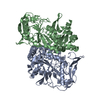
 Movie
Movie Controller


+ Open data

- Basic information
| Entry | Database: PDB / ID: 1j4b | ||||||
|---|---|---|---|---|---|---|---|
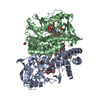
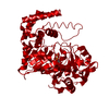
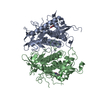
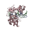
| Title | Recombinant Mouse-Muscle Adenylosuccinate Synthetase | ||||||
 Components Components | adenylosuccinate synthetase Adenylosuccinate synthase Adenylosuccinate synthase | ||||||
 Keywords Keywords | LIGASE / GTP-hydrolyzing enzymes / purine nucleotide cycle | ||||||
| Function / homology |  Function and homology information Function and homology informationPurine ribonucleoside monophosphate biosynthesis / AMP biosynthetic process / adenylosuccinate synthase / adenylosuccinate synthase activity / purine nucleotide metabolic process / aspartate metabolic process / IMP metabolic process / 'de novo' AMP biosynthetic process / AMP salvage / actin filament binding ...Purine ribonucleoside monophosphate biosynthesis / AMP biosynthetic process / adenylosuccinate synthase / adenylosuccinate synthase activity / purine nucleotide metabolic process / aspartate metabolic process / IMP metabolic process / 'de novo' AMP biosynthetic process / AMP salvage / actin filament binding / GTPase activity / GTP binding / magnesium ion binding / membrane / identical protein binding / cytoplasmSimilarity search - Function | ||||||
| Biological species |  Mus musculus (house mouse) Mus musculus (house mouse) | ||||||
| Method | X-RAY DIFFRACTION / SYNCHROTRON / MOLECULAR REPLACEMENT / Resolution: 2.5 Å | ||||||
 Authors Authors | Iancu, C.V. / Borza, T. / Choe, J.Y. / Fromm, H.J. / Honzatko, R.B. | ||||||
 Citation Citation | Journal: J.Biol.Chem. / Year: 2001 Title: Recombinant mouse muscle adenylosuccinate synthetase: overexpression, kinetics, and crystal structure. Authors: Iancu, C.V. / Borza, T. / Choe, J.Y. / Fromm, H.J. / Honzatko, R.B. | ||||||
| History |
|
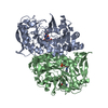
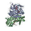
- Structure visualization
| Structure viewer | Molecule: MolmilJmol/JSmol |
|---|
- Downloads & links
-Download
| PDBx/mmCIF format | 1j4b.cif.gz | 97.5 KB | Display | PDBx/mmCIF format |
|---|---|---|---|---|
| PDB format | pdb1j4b.ent.gz | 73.9 KB | Display | PDB format |
| PDBx/mmJSON format | 1j4b.json.gz | Tree view | PDBx/mmJSON format | |
| Others |  Other downloads Other downloads |
-Validation report
| Arichive directory | https://data.pdbj.org/pub/pdb/validation_reports/j4/1j4bftp://data.pdbj.org/pub/pdb/validation_reports/j4/1j4b | HTTPS FTP |
|---|
-Related structure data
| Related structure data |  1sonS S: Starting model for refinement |
|---|---|
| Similar structure data |


-Links
 PDBj
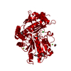
PDBj- Assembly
| Deposited unit | 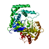
| ||||||||
|---|---|---|---|---|---|---|---|---|---|
| 1 | 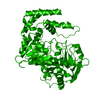
| ||||||||
| Unit cell |
|
-Components
| #1: Protein | Adenylosuccinate synthase / AdSS1 Mass: 50321.301 Da / Num. of mol.: 1 Source method: isolated from a genetically manipulated source Source: (gene. exp.) Mus musculus (house mouse) / Tissue: muscleSkeletal muscle / Cellular location: cytoplasm / Gene: Adss1 / Plasmid: pET28b / Species (production host): Escherichia coli / Cellular location (production host): cytoplasm / Production host:  Escherichia coli BL21(DE3) (bacteria) / Strain (production host): BL21 (DE3) / References: UniProt: P28650, adenylosuccinate synthase Escherichia coli BL21(DE3) (bacteria) / Strain (production host): BL21 (DE3) / References: UniProt: P28650, adenylosuccinate synthase |
|---|---|
| #2: Water | ChemComp-HOH / Water Mass: 18.015 Da / Num. of mol.: 96 / Source method: isolated from a natural source / Formula: H2O Mass: 18.015 Da / Num. of mol.: 96 / Source method: isolated from a natural source / Formula: H2O |
-Experimental details
-Experiment
| Experiment | Method: X-RAY DIFFRACTION / Number of used crystals: 1 |
|---|
- Sample preparation
| Crystal | Density Matthews: 2.4 Å3/Da / Density % sol: 48 % | |||||||||||||||||||||||||||||||||||||||||||||||||||||||||||||||
|---|---|---|---|---|---|---|---|---|---|---|---|---|---|---|---|---|---|---|---|---|---|---|---|---|---|---|---|---|---|---|---|---|---|---|---|---|---|---|---|---|---|---|---|---|---|---|---|---|---|---|---|---|---|---|---|---|---|---|---|---|---|---|---|---|
| Crystal grow | pH: 6.5 Details: Cacodylate 100 mM, pH6.5 PEG 8000 18% 200 mM Calcium Acetate | |||||||||||||||||||||||||||||||||||||||||||||||||||||||||||||||
| Crystal grow | *PLUS Temperature: 22 ℃ / pH: 7.5 / Method: vapor diffusion, hanging drop | |||||||||||||||||||||||||||||||||||||||||||||||||||||||||||||||
| Components of the solutions | *PLUS
|
-Data collection
| Diffraction | Mean temperature: 100 K |
|---|---|
| Diffraction source | Source: SYNCHROTRON / Site: SSRL  / Beamline: BL9-2 / Wavelength: 0.979 / Beamline: BL9-2 / Wavelength: 0.979 |
| Detector | Type: ADSC QUANTUM 4 / Detector: CCD / Date: Feb 1, 2001 |
| Radiation | Protocol: SINGLE WAVELENGTH / Monochromatic (M) / Laue (L): M / Scattering type: x-ray |
| Radiation wavelength | Wavelength: 0.979 Å / Relative weight: 1 |
| Reflection | Resolution: 2.5→39.6 Å / Num. obs: 16066 / % possible obs: 90.3 % / Observed criterion σ(I): 0 / Redundancy: 8.3 % / Biso Wilson estimate: 27.4 Å2 / Rmerge(I) obs: 0.063 / Rsym value: 0.063 / Net I/σ(I): 24 |
| Reflection shell | Resolution: 2.5→2.59 Å / Redundancy: 4 % / Rmerge(I) obs: 0.224 / Mean I/σ(I) obs: 2 / Rsym value: 0.224 / % possible all: 60 |
| Reflection | *PLUS Num. measured all: 132963 |
| Reflection shell | *PLUS % possible obs: 60 % |
- Processing
| Software |
| ||||||||||||||||||||||||||||||||||||||||||||||||||||||||||||
|---|---|---|---|---|---|---|---|---|---|---|---|---|---|---|---|---|---|---|---|---|---|---|---|---|---|---|---|---|---|---|---|---|---|---|---|---|---|---|---|---|---|---|---|---|---|---|---|---|---|---|---|---|---|---|---|---|---|---|---|---|---|
| Refinement | Method to determine structure: MOLECULAR REPLACEMENT Starting model: 1SON Resolution: 2.5→39.61 Å / Rfactor Rfree error: 0.007 / Data cutoff high absF: 132841.62 / Data cutoff low absF: 0 / Isotropic thermal model: RESTRAINED / Cross valid method: THROUGHOUT / σ(F): 0
| ||||||||||||||||||||||||||||||||||||||||||||||||||||||||||||
| Solvent computation | Solvent model: FLAT MODEL / Bsol: 34.0352 Å2 / ksol: 0.357928 e/Å3 | ||||||||||||||||||||||||||||||||||||||||||||||||||||||||||||
| Displacement parameters | Biso mean: 40.8 Å2
| ||||||||||||||||||||||||||||||||||||||||||||||||||||||||||||
| Refine analyze |
| ||||||||||||||||||||||||||||||||||||||||||||||||||||||||||||
| Refinement step | Cycle: LAST / Resolution: 2.5→39.61 Å
| ||||||||||||||||||||||||||||||||||||||||||||||||||||||||||||
| Refine LS restraints |
| ||||||||||||||||||||||||||||||||||||||||||||||||||||||||||||
| LS refinement shell | Resolution: 2.5→2.66 Å / Rfactor Rfree error: 0.026 / Total num. of bins used: 6
| ||||||||||||||||||||||||||||||||||||||||||||||||||||||||||||
| Xplor file |
| ||||||||||||||||||||||||||||||||||||||||||||||||||||||||||||
| Software | *PLUS Name: CNS / Version: 1 / Classification: refinement | ||||||||||||||||||||||||||||||||||||||||||||||||||||||||||||
| Refinement | *PLUS σ(F): 0 / % reflection Rfree: 9.9 % | ||||||||||||||||||||||||||||||||||||||||||||||||||||||||||||
| Solvent computation | *PLUS | ||||||||||||||||||||||||||||||||||||||||||||||||||||||||||||
| Displacement parameters | *PLUS Biso mean: 40.8 Å2 | ||||||||||||||||||||||||||||||||||||||||||||||||||||||||||||
| Refine LS restraints | *PLUS
| ||||||||||||||||||||||||||||||||||||||||||||||||||||||||||||
| LS refinement shell | *PLUS Rfactor Rfree: 0.336 / % reflection Rfree: 9.3 % / Rfactor Rwork: 0.265 |
