 Movie
Movie Controller

[English] 日本語
 Yorodumi
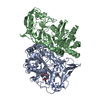
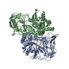
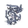
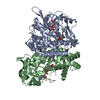
- PDB-1adi: STRUCTURE OF ADENYLOSUCCINATE SYNTHETASE AT PH 6.5 AND 25 DEGREES... -
+ Open data

- Basic information
| Entry | Database: PDB / ID: 1adi | ||||||
|---|---|---|---|---|---|---|---|
| Title | STRUCTURE OF ADENYLOSUCCINATE SYNTHETASE AT PH 6.5 AND 25 DEGREES CELSIUS | ||||||
 Components Components | ADENYLOSUCCINATE SYNTHETASE Adenylosuccinate synthase Adenylosuccinate synthase | ||||||
 Keywords Keywords | LIGASE / PURINE NUCLEOTIDE BIOSYNTHESIS / GTP-HYDROLYZING ENZYME | ||||||
| Function / homology |  Function and homology informationadenylosuccinate synthase / adenylosuccinate synthase activity / adenosine biosynthetic process / IMP metabolic process / 'de novo' AMP biosynthetic process / nucleobase-containing small molecule interconversion / purine nucleotide biosynthetic process / guanosine tetraphosphate binding / DNA damage response / GTP binding ...adenylosuccinate synthase / adenylosuccinate synthase activity / adenosine biosynthetic process / IMP metabolic process / 'de novo' AMP biosynthetic process / nucleobase-containing small molecule interconversion / purine nucleotide biosynthetic process / guanosine tetraphosphate binding / DNA damage response / GTP binding / magnesium ion binding / membrane / cytosol / cytoplasm Function and homology informationadenylosuccinate synthase / adenylosuccinate synthase activity / adenosine biosynthetic process / IMP metabolic process / 'de novo' AMP biosynthetic process / nucleobase-containing small molecule interconversion / purine nucleotide biosynthetic process / guanosine tetraphosphate binding / DNA damage response / GTP binding ...adenylosuccinate synthase / adenylosuccinate synthase activity / adenosine biosynthetic process / IMP metabolic process / 'de novo' AMP biosynthetic process / nucleobase-containing small molecule interconversion / purine nucleotide biosynthetic process / guanosine tetraphosphate binding / DNA damage response / GTP binding / magnesium ion binding / membrane / cytosol / cytoplasmSimilarity search - Function | ||||||
| Biological species |  Escherichia coli (E. coli) Escherichia coli (E. coli) | ||||||
| Method | X-RAY DIFFRACTION / Resolution: 2.5 Å | ||||||
 Authors Authors | Silva, M.M. / Poland, B.W. / Hoffman, C.M. / Fromm, H.J. / Honzatko, R.B. | ||||||
 Citation Citation | Journal: J.Mol.Biol. / Year: 1995 Title: Refined crystal structures of unligated adenylosuccinate synthetase from Escherichia coli. Authors: Silva, M.M. / Poland, B.W. / Hoffman, C.R. / Fromm, H.J. / Honzatko, R.B. #1: Journal: J.Biol.Chem. / Year: 1993Title: Crystal Structure of Adenylosuccinate Synthetase from Escherichia Coli. Evidence for Convergent Evolution of GTP-Binding Domains Authors: Poland, B.W. / Silva, M.M. / Serra, M.A. / Cho, Y. / Kim, K.H. / Harris, E.M. / Honzatko, R.B. | ||||||
| History |
|
- Structure visualization
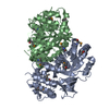
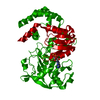
| Structure viewer | Molecule: MolmilJmol/JSmol |
|---|
- Downloads & links
-Download
| PDBx/mmCIF format | 1adi.cif.gz | 232 KB | Display | PDBx/mmCIF format |
|---|---|---|---|---|
| PDB format | pdb1adi.ent.gz | 189.6 KB | Display | PDB format |
| PDBx/mmJSON format | 1adi.json.gz | Tree view | PDBx/mmJSON format | |
| Others |  Other downloads Other downloads |
-Validation report
| Arichive directory | https://data.pdbj.org/pub/pdb/validation_reports/ad/1adiftp://data.pdbj.org/pub/pdb/validation_reports/ad/1adi | HTTPS FTP |
|---|
-Related structure data


-Links
 PDBj
PDBj- Assembly
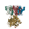
| Deposited unit | 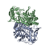
| ||||||||
|---|---|---|---|---|---|---|---|---|---|
| 1 |
| ||||||||
| Unit cell |
| ||||||||
| Noncrystallographic symmetry (NCS) | NCS oper: (Code: given Matrix: (-0.98902, 0.01348, -0.14717), Vector : |
-Components
| #1: Protein | Adenylosuccinate synthase Mass: 47269.598 Da / Num. of mol.: 2 / Source method: isolated from a natural source / Source: (natural) Escherichia coli (E. coli)Strain: PUR A STRAIN H1238 (A GIFT FROM DR. B. BACHMAN (GENETIC CENTER, YALE UNIVERSITY)) References: UniProt: P0A7D4, adenylosuccinate synthase#2: Water | ChemComp-HOH / | Water Mass: 18.015 Da / Num. of mol.: 439 / Source method: isolated from a natural source / Formula: H2O Mass: 18.015 Da / Num. of mol.: 439 / Source method: isolated from a natural source / Formula: H2O |
|---|
-Experimental details
-Experiment
| Experiment | Method: X-RAY DIFFRACTION |
|---|
- Sample preparation
| Crystal | Density Matthews: 2.17 Å3/Da / Density % sol: 43.34 % | ||||||||||||||||||||
|---|---|---|---|---|---|---|---|---|---|---|---|---|---|---|---|---|---|---|---|---|---|
| Crystal grow | *PLUS pH: 6.5 / Method: vapor diffusion, hanging drop | ||||||||||||||||||||
| Components of the solutions | *PLUS
|
-Data collection
| Diffraction source | Wavelength: 1.5418 |
|---|---|
| Detector | Type: SIEMENS-NICOLET X100 / Detector: AREA DETECTOR |
| Radiation | Monochromatic (M) / Laue (L): M / Scattering type: x-ray |
| Radiation wavelength | Wavelength: 1.5418 Å / Relative weight: 1 |
| Reflection | Resolution: 2.5→15 Å / Num. obs: 104085 / % possible obs: 99 % / Observed criterion σ(I): 2 / Redundancy: 3.24 % / Rmerge(I) obs: 0.06 |
| Reflection | *PLUS Num. obs: 32170 / Num. measured all: 104085 |
- Processing
| Software |
| ||||||||||||||||||||||||||||||||||||||||||||||||||||||||||||
|---|---|---|---|---|---|---|---|---|---|---|---|---|---|---|---|---|---|---|---|---|---|---|---|---|---|---|---|---|---|---|---|---|---|---|---|---|---|---|---|---|---|---|---|---|---|---|---|---|---|---|---|---|---|---|---|---|---|---|---|---|---|
| Refinement | Resolution: 2.5→5 Å / σ(F): 0 Details: THERE ARE TWO REGIONS THAT ARE DISORDERED CONSISTING OF RESIDUES 121 - 130 AND 298 - 303.
| ||||||||||||||||||||||||||||||||||||||||||||||||||||||||||||
| Displacement parameters | Biso mean: 25.5 Å2 | ||||||||||||||||||||||||||||||||||||||||||||||||||||||||||||
| Refine analyze | Luzzati coordinate error obs: 0.25 Å | ||||||||||||||||||||||||||||||||||||||||||||||||||||||||||||
| Refinement step | Cycle: LAST / Resolution: 2.5→5 Å
| ||||||||||||||||||||||||||||||||||||||||||||||||||||||||||||
| Refine LS restraints |
| ||||||||||||||||||||||||||||||||||||||||||||||||||||||||||||
| Software | *PLUS Name: 'X-PLOR' / Classification: refinement | ||||||||||||||||||||||||||||||||||||||||||||||||||||||||||||
| Refine LS restraints | *PLUS
|
