Mass: 18.015 Da / Num. of mol.: 585 / Source method: isolated from a natural source / Formula: H2O
-
Details
Sequence details
NATIVE SEQUENCE HAS AN N-TERMINAL HIS-TAG AND THROMBIN SITE ADDED
-
Experimental details
-
Experiment
Experiment
Method: X-RAY DIFFRACTION / Number of used crystals: 1
-
Sample preparation
Crystal
Density Matthews: 2.71 Å3/Da / Density % sol: 54.6 % / Description: NONE
Crystal grow
pH: 7 Details: PROTEIN WAS AT 8 MG/ML AND 200 NL PLUS 200 NL DROPS WERE SET UP WITH THE RESERVOIR CONDITION OF 10% PEG 4000, 0.2 M MGCL2, 0.1 M TRIS PH 7.
Resolution: 1.33→89.47 Å / Cor.coef. Fo:Fc: 0.984 / Cor.coef. Fo:Fc free: 0.98 / SU B: 1.151 / SU ML: 0.021 / Cross valid method: THROUGHOUT / ESU R: 0.036 / ESU R Free: 0.034 / Stereochemistry target values: MAXIMUM LIKELIHOOD Details: HYDROGENS HAVE BEEN ADDED IN THE RIDING POSITIONS. U VALUES REFINED INDIVIDUALLY THE PLP COFACTOR IS NOT WELL RESOLVED IN THE DENSITY.
Rfactor
Num. reflection
% reflection
Selection details
Rfree
0.12644
5822
5 %
RANDOM
Rwork
0.107
-
-
-
obs
0.10797
110474
99.81 %
-
Solvent computation
Ion probe radii: 0.8 Å / Shrinkage radii: 0.8 Å / VDW probe radii: 1.2 Å / Solvent model: MASK
 Movie
Movie Controller

 Yorodumi
 Open data
 Basic information
 Components
 Keywords
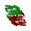
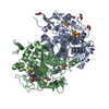
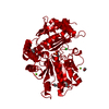
 TRANSFERASE /
  Function and homology information
 Authors
 Citation
 Structure visualization
 Downloads & links
 Other downloads


 PDBj
PDBj


 Assembly


 Mass: 24.305 Da / Num. of mol.: 2 / Source method: obtained synthetically / Formula: Mg
 Mass: 46.068 Da / Num. of mol.: 2 / Source method: obtained synthetically / Formula: C2H6O
 Mass: 92.094 Da / Num. of mol.: 3 / Source method: obtained synthetically / Formula: C3H8O3
 Mass: 62.068 Da / Num. of mol.: 2 / Source method: obtained synthetically / Formula: C2H6O2
 Mass: 247.142 Da / Num. of mol.: 1 / Source method: obtained synthetically / Formula: C8H10NO6P
 Mass: 35.453 Da / Num. of mol.: 2 / Source method: obtained synthetically / Formula: Cl
 Sample preparation
 / Beamline: MX2 / Wavelength: 0.9184
  Processing