 Movie
Movie Controller

[English] 日本語
 Yorodumi
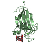
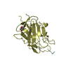
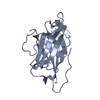
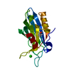
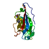
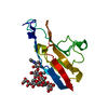
- PDB-1ixl: Crystal structure of uncharacterized protein PH1136 from Pyrococc... -
+ Open data

- Basic information
| Entry | Database: PDB / ID: 1ixl | ||||||
|---|---|---|---|---|---|---|---|
| Title | Crystal structure of uncharacterized protein PH1136 from Pyrococcus horikoshii | ||||||
 Components Components | hypothetical protein PH1136 Hypothesis Hypothesis | ||||||
 Keywords Keywords | STRUCTURAL GENOMICS / UNKNOWN FUNCTION / alpha+beta / hot-dog-fold | ||||||
| Function / homology | Thioesterase domain / Thioesterase superfamily / Hotdog Thioesterase / Thiol Ester Dehydrase; Chain A / HotDog domain superfamily / Roll / Alpha Beta / 4HBT domain-containing protein Function and homology information Function and homology information | ||||||
| Biological species |   Pyrococcus horikoshii (archaea) Pyrococcus horikoshii (archaea) | ||||||
| Method | X-RAY DIFFRACTION / SYNCHROTRON / MAD / Resolution: 1.94 Å | ||||||
 Authors Authors | Tajika, Y. / Sakai, N. / Tanaka, Y. / Yao, M. / Watanabe, N. / Tanaka, I. | ||||||
 Citation Citation | Journal: Proteins / Year: 2004 Title: Crystal structure of conserved protein PH1136 from Pyrococcus horikoshii. Authors: Tajika, Y. / Sakai, N. / Tanaka, Y. / Yao, M. / Watanabe, N. / Tanaka, I. | ||||||
| History |
|
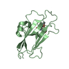
- Structure visualization
| Structure viewer | Molecule: MolmilJmol/JSmol |
|---|
- Downloads & links
-Download
| PDBx/mmCIF format | 1ixl.cif.gz | 39.1 KB | Display | PDBx/mmCIF format |
|---|---|---|---|---|
| PDB format | pdb1ixl.ent.gz | 27.1 KB | Display | PDB format |
| PDBx/mmJSON format | 1ixl.json.gz | Tree view | PDBx/mmJSON format | |
| Others |  Other downloads Other downloads |
-Validation report
| Arichive directory | https://data.pdbj.org/pub/pdb/validation_reports/ix/1ixlftp://data.pdbj.org/pub/pdb/validation_reports/ix/1ixl | HTTPS FTP |
|---|
-Related structure data
| Similar structure data |
|---|


-Links
 PDBj
PDBj
- Assembly
| Deposited unit | 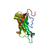
| ||||||||
|---|---|---|---|---|---|---|---|---|---|
| 1 |
| ||||||||
| 2 | 
| ||||||||
| Unit cell |
|
-Components
| #1: Protein | Hypothesis Mass: 14893.058 Da / Num. of mol.: 1 Source method: isolated from a genetically manipulated source Source: (gene. exp.) Pyrococcus horikoshii (archaea) / Plasmid: pET-22b / Species (production host): Escherichia coli / Production host:  Escherichia coli BL21(DE3) (bacteria) / Strain (production host): BL21 (DE3) / References: UniProt: O58863 Escherichia coli BL21(DE3) (bacteria) / Strain (production host): BL21 (DE3) / References: UniProt: O58863 |
|---|---|
| #2: Water | ChemComp-HOH / Water Mass: 18.015 Da / Num. of mol.: 89 / Source method: isolated from a natural source / Formula: H2O Mass: 18.015 Da / Num. of mol.: 89 / Source method: isolated from a natural source / Formula: H2O |
-Experimental details
-Experiment
| Experiment | Method: X-RAY DIFFRACTION / Number of used crystals: 1 |
|---|
- Sample preparation
| Crystal | Density Matthews: 2.58 Å3/Da / Density % sol: 52.3 % | ||||||||||||||||||||||||||||||
|---|---|---|---|---|---|---|---|---|---|---|---|---|---|---|---|---|---|---|---|---|---|---|---|---|---|---|---|---|---|---|---|
| Crystal grow | Temperature: 293 K / Method: vapor diffusion, hanging drop / pH: 8.2 Details: Tris-HCl, Ammonium Sulfate, pH 8.2, VAPOR DIFFUSION, HANGING DROP, temperature 293K | ||||||||||||||||||||||||||||||
| Crystal grow | *PLUS Temperature: 4 ℃ / pH: 8.5 / Method: vapor diffusion | ||||||||||||||||||||||||||||||
| Components of the solutions | *PLUS
|
-Data collection
| Diffraction | Mean temperature: 100 K | ||||||||||||
|---|---|---|---|---|---|---|---|---|---|---|---|---|---|
| Diffraction source | Source: SYNCHROTRON / Site: SPring-8  / Beamline: BL45PX / Wavelength: 0.9000, 0.9785, 0.9791 / Beamline: BL45PX / Wavelength: 0.9000, 0.9785, 0.9791 | ||||||||||||
| Detector | Type: RIGAKU / Detector: IMAGE PLATE / Date: Mar 4, 2002 | ||||||||||||
| Radiation | Monochromator: MIRRORS + Si (111) / Protocol: MAD / Monochromatic (M) / Laue (L): M / Scattering type: x-ray | ||||||||||||
| Radiation wavelength |
| ||||||||||||
| Reflection | Resolution: 1.94→31 Å / Num. obs: 11527 / % possible obs: 96.8 % / Observed criterion σ(I): -3 / Redundancy: 8.23 % / Biso Wilson estimate: 23.6 Å2 / Rmerge(I) obs: 0.083 / Net I/σ(I): 8.1 | ||||||||||||
| Reflection shell | Resolution: 1.94→2.01 Å / Redundancy: 8.39 % / Rmerge(I) obs: 0.33 / Mean I/σ(I) obs: 3.4 / Num. unique all: 1174 / % possible all: 100 | ||||||||||||
| Reflection | *PLUS Lowest resolution: 50 Å | ||||||||||||
| Reflection shell | *PLUS % possible obs: 100 % / Rmerge(I) obs: 0.332 |
- Processing
| Software |
| ||||||||||||||||||||||||||||||||
|---|---|---|---|---|---|---|---|---|---|---|---|---|---|---|---|---|---|---|---|---|---|---|---|---|---|---|---|---|---|---|---|---|---|
| Refinement | Method to determine structure: MAD / Resolution: 1.94→10 Å / Isotropic thermal model: Isotropic / Cross valid method: THROUGHOUT / σ(F): 0 / σ(I): 0 / Stereochemistry target values: Engh & Huber
| ||||||||||||||||||||||||||||||||
| Solvent computation | Solvent model: throughput / Bsol: 49.8 Å2 / ksol: 0.462 e/Å3 | ||||||||||||||||||||||||||||||||
| Displacement parameters | Biso mean: 30.7 Å2
| ||||||||||||||||||||||||||||||||
| Refine analyze |
| ||||||||||||||||||||||||||||||||
| Refinement step | Cycle: LAST / Resolution: 1.94→10 Å
| ||||||||||||||||||||||||||||||||
| Refine LS restraints |
| ||||||||||||||||||||||||||||||||
| LS refinement shell | Refine-ID: X-RAY DIFFRACTION / Total num. of bins used: 10
| ||||||||||||||||||||||||||||||||
| Xplor file | Serial no: 1 / Param file: protein_rep.param / Topol file: protein.top | ||||||||||||||||||||||||||||||||
| Refinement | *PLUS Lowest resolution: 50 Å | ||||||||||||||||||||||||||||||||
| Solvent computation | *PLUS | ||||||||||||||||||||||||||||||||
| Displacement parameters | *PLUS | ||||||||||||||||||||||||||||||||
| Refine LS restraints | *PLUS
|
