 Movie
Movie Controller


+ Open data

- Basic information
| Entry | Database: PDB / ID: 1o7v | ||||||||||||
|---|---|---|---|---|---|---|---|---|---|---|---|---|---|
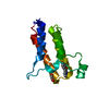
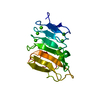
| Title | High resolution structure of Siglec-7 | ||||||||||||
 Components Components | SIALIC ACID BINDING IG-LIKE LECTIN 7 | ||||||||||||
 Keywords Keywords |  CELL ADHESION / SIGLEC / IMMUNOLOGLOBULIN-LIKE FOLD / LECTIN / SIALIC ACID BINDING PROTEIN / IMMUNE SYSTEM CELL ADHESION / SIGLEC / IMMUNOLOGLOBULIN-LIKE FOLD / LECTIN / SIALIC ACID BINDING PROTEIN / IMMUNE SYSTEM | ||||||||||||
| Function / homology |  Function and homology informationsialic acid binding / Immunoregulatory interactions between a Lymphoid and a non-Lymphoid cell / signaling receptor activity / carbohydrate binding / cell adhesion / plasma membrane Function and homology informationsialic acid binding / Immunoregulatory interactions between a Lymphoid and a non-Lymphoid cell / signaling receptor activity / carbohydrate binding / cell adhesion / plasma membraneSimilarity search - Function | ||||||||||||
| Biological species |  Homo sapiens (human) Homo sapiens (human) | ||||||||||||
| Method | X-RAY DIFFRACTION / SYNCHROTRON / MOLECULAR REPLACEMENT / Resolution: 1.9 Å | ||||||||||||
 Authors Authors | Alphey, M.S. / Attrill, H. / Crocker, P.R. / Van Aalten, D.M.F. | ||||||||||||
 Citation Citation | Journal: J.Biol.Chem. / Year: 2003 Title: High resolution crystal structures of Siglec-7. Insights into ligand specificity in the Siglec family. Authors: Alphey, M.S. / Attrill, H. / Crocker, P.R. / van Aalten, D.M. | ||||||||||||
| History |
| ||||||||||||
| Remark 700 | SHEET THE SHEET STRUCTURE OF THIS MOLECULE IS BIFURCATED. IN ORDER TO REPRESENT THIS FEATURE IN ... SHEET THE SHEET STRUCTURE OF THIS MOLECULE IS BIFURCATED. IN ORDER TO REPRESENT THIS FEATURE IN THE SHEET RECORDS BELOW, TWO SHEETS ARE DEFINED. |
- Structure visualization
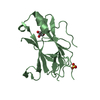
| Structure viewer | Molecule: MolmilJmol/JSmol |
|---|
- Downloads & links
-Download
| PDBx/mmCIF format | 1o7v.cif.gz | 46.3 KB | Display | PDBx/mmCIF format |
|---|---|---|---|---|
| PDB format | pdb1o7v.ent.gz | 30.5 KB | Display | PDB format |
| PDBx/mmJSON format | 1o7v.json.gz | Tree view | PDBx/mmJSON format | |
| Others |  Other downloads Other downloads |
-Validation report
| Arichive directory | https://data.pdbj.org/pub/pdb/validation_reports/o7/1o7vftp://data.pdbj.org/pub/pdb/validation_reports/o7/1o7v | HTTPS FTP |
|---|
-Related structure data
| Related structure data |  1o7sSC S: Starting model for refinement C: citing same article ( |
|---|---|
| Similar structure data |


-Links
 PDBj
PDBj
- Assembly
| Deposited unit | 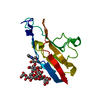
| ||||||||
|---|---|---|---|---|---|---|---|---|---|
| 1 |
| ||||||||
| Unit cell |
|
-Components
| #1: Protein | Mass: 14655.292 Da / Num. of mol.: 1 / Fragment: V-SET SIALIC ACID BINDING DOMAIN, RESIDUES 18-144 Source method: isolated from a genetically manipulated source Source: (gene. exp.) Homo sapiens (human) / Plasmid: PDEF / Cell line (production host): CHO LEC1 / Production host:   Cricetulus griseus (Chinese hamster) / References: UniProt: Q9Y286 Cricetulus griseus (Chinese hamster) / References: UniProt: Q9Y286 |
|---|---|
| #2: Polysaccharide | alpha-D-mannopyranose-(1-3)-[alpha-D-mannopyranose-(1-6)]alpha-D-mannopyranose-(1-6)-[alpha-D- ...alpha-D-mannopyranose-(1-3)-[alpha-D-mannopyranose-(1-6)]alpha-D-mannopyranose-(1-6)-[alpha-D-mannopyranose-(1-3)]beta-D-mannopyranose-(1-4)-2-acetamido-2-deoxy-beta-D-glucopyranose-(1-4)-2-acetamido-2-deoxy-beta-D-glucopyranose / Mass: 1235.105 Da / Num. of mol.: 1 Source method: isolated from a genetically manipulated source |
| #3: Water | ChemComp-HOH / Water Mass: 18.015 Da / Num. of mol.: 86 / Source method: isolated from a natural source / Formula: H2O Mass: 18.015 Da / Num. of mol.: 86 / Source method: isolated from a natural source / Formula: H2O |
-Experimental details
-Experiment
| Experiment | Method: X-RAY DIFFRACTION / Number of used crystals: 1 |
|---|
- Sample preparation
| Crystal | Density Matthews: 1.731 Å3/Da / Density % sol: 27.63 % | ||||||||||||||||||||||||||||||||||||||||||||||||||||||
|---|---|---|---|---|---|---|---|---|---|---|---|---|---|---|---|---|---|---|---|---|---|---|---|---|---|---|---|---|---|---|---|---|---|---|---|---|---|---|---|---|---|---|---|---|---|---|---|---|---|---|---|---|---|---|---|
| Crystal grow | Temperature: 293 K / Method: vapor diffusion, hanging drop / pH: 6.5 Details: 0.2 M SODIUM ACETATE TRIHYDRATE 0.1 M SODIUM CACODYLATE PH 6.5, 30 % PEG 8000 | ||||||||||||||||||||||||||||||||||||||||||||||||||||||
| Crystal grow | *PLUS Temperature: 20 ℃ / pH: 8 / Method: vapor diffusion, hanging drop | ||||||||||||||||||||||||||||||||||||||||||||||||||||||
| Components of the solutions | *PLUS
|
-Data collection
| Diffraction | Mean temperature: 100 K |
|---|---|
| Diffraction source | Source: SYNCHROTRON / Site: ESRF  / Beamline: ID29 / Wavelength: 0.933 / Beamline: ID29 / Wavelength: 0.933 |
| Detector | Type: ADSC CCD / Detector: CCD / Date: Oct 29, 2001 |
| Radiation | Protocol: SINGLE WAVELENGTH / Monochromatic (M) / Laue (L): M / Scattering type: x-ray |
| Radiation wavelength | Wavelength: 0.933 Å / Relative weight: 1 |
| Reflection | Resolution: 1.9→25 Å / Num. obs: 9146 / % possible obs: 94.8 % / Redundancy: 3.4 % / Biso Wilson estimate: 17.3 Å2 / Rmerge(I) obs: 0.061 / Net I/σ(I): 11.7 |
| Reflection shell | Resolution: 1.9→1.97 Å / Redundancy: 2.9 % / Rmerge(I) obs: 0.312 / Mean I/σ(I) obs: 2.4 / % possible all: 96.3 |
| Reflection | *PLUS Lowest resolution: 25 Å / Num. obs: 9206 / Num. measured all: 31409 |
| Reflection shell | *PLUS % possible obs: 96.3 % |
- Processing
| Software |
| ||||||||||||||||||||||||||||||||||||||||||||||||||||||||||||||||||||||||||||||||
|---|---|---|---|---|---|---|---|---|---|---|---|---|---|---|---|---|---|---|---|---|---|---|---|---|---|---|---|---|---|---|---|---|---|---|---|---|---|---|---|---|---|---|---|---|---|---|---|---|---|---|---|---|---|---|---|---|---|---|---|---|---|---|---|---|---|---|---|---|---|---|---|---|---|---|---|---|---|---|---|---|---|
| Refinement | Method to determine structure: MOLECULAR REPLACEMENT Starting model: PDB ENTRY 1O7S Resolution: 1.9→24.25 Å / Rfactor Rfree error: 0.012 / Data cutoff high absF: 1412842.78 / Cross valid method: THROUGHOUT / σ(F): 0 Details: FULL N-LINKED GLYCAN STRUCTURE SEEN (HIGH MANNOSE STRUCTURE)
| ||||||||||||||||||||||||||||||||||||||||||||||||||||||||||||||||||||||||||||||||
| Displacement parameters | Biso mean: 34.09 Å2
| ||||||||||||||||||||||||||||||||||||||||||||||||||||||||||||||||||||||||||||||||
| Refine analyze |
| ||||||||||||||||||||||||||||||||||||||||||||||||||||||||||||||||||||||||||||||||
| Refinement step | Cycle: LAST / Resolution: 1.9→24.25 Å
| ||||||||||||||||||||||||||||||||||||||||||||||||||||||||||||||||||||||||||||||||
| Refine LS restraints |
| ||||||||||||||||||||||||||||||||||||||||||||||||||||||||||||||||||||||||||||||||
| LS refinement shell | Resolution: 1.9→1.99 Å / Rfactor Rfree error: 0.035 / Total num. of bins used: 8
| ||||||||||||||||||||||||||||||||||||||||||||||||||||||||||||||||||||||||||||||||
| Xplor file |
| ||||||||||||||||||||||||||||||||||||||||||||||||||||||||||||||||||||||||||||||||
| Refinement | *PLUS Highest resolution: 1.9 Å / Lowest resolution: 25 Å / Rfactor Rfree: 0.255 / Rfactor Rwork: 0.21 | ||||||||||||||||||||||||||||||||||||||||||||||||||||||||||||||||||||||||||||||||
| Solvent computation | *PLUS | ||||||||||||||||||||||||||||||||||||||||||||||||||||||||||||||||||||||||||||||||
| Displacement parameters | *PLUS | ||||||||||||||||||||||||||||||||||||||||||||||||||||||||||||||||||||||||||||||||
| Refine LS restraints | *PLUS Type: c_bond_d / Dev ideal: 0.007 |
