 Movie
Movie Controller

[English] 日本語
 Yorodumi
- PDB-1il0: X-RAY CRYSTAL STRUCTURE OF THE E170Q MUTANT OF HUMAN L-3-HYDROXYA... -
+ Open data

- Basic information
| Entry | Database: PDB / ID: 1il0 | ||||||
|---|---|---|---|---|---|---|---|
| Title | X-RAY CRYSTAL STRUCTURE OF THE E170Q MUTANT OF HUMAN L-3-HYDROXYACYL-COA DEHYDROGENASE | ||||||
 Components Components | 3-hydroxyacyl-CoA dehydrogenase | ||||||
 Keywords Keywords | OXIDOREDUCTASE / ABORTIVE TERNARY COMPLEX | ||||||
| Function / homology |  Function and homology information Function and homology informationBeta oxidation of lauroyl-CoA to decanoyl-CoA-CoA / Beta oxidation of butanoyl-CoA to acetyl-CoA / Beta oxidation of hexanoyl-CoA to butanoyl-CoA / Beta oxidation of octanoyl-CoA to hexanoyl-CoA / Beta oxidation of decanoyl-CoA to octanoyl-CoA-CoA / 3-hydroxyacyl-CoA dehydrogenase / 3-hydroxyacyl-CoA dehydrogenase activity / fatty acid beta-oxidation / regulation of insulin secretion / NAD+ binding ...Beta oxidation of lauroyl-CoA to decanoyl-CoA-CoA / Beta oxidation of butanoyl-CoA to acetyl-CoA / Beta oxidation of hexanoyl-CoA to butanoyl-CoA / Beta oxidation of octanoyl-CoA to hexanoyl-CoA / Beta oxidation of decanoyl-CoA to octanoyl-CoA-CoA / 3-hydroxyacyl-CoA dehydrogenase / 3-hydroxyacyl-CoA dehydrogenase activity / fatty acid beta-oxidation / regulation of insulin secretion / NAD+ binding / negative regulation of insulin secretion / Mitochondrial protein degradation / response to activity / response to insulin / positive regulation of cold-induced thermogenesis / transferase activity / mitochondrial matrix / response to xenobiotic stimulus / mitochondrion / nucleoplasm / identical protein binding / cytoplasmSimilarity search - Function | ||||||
| Biological species |  Homo sapiens (human) Homo sapiens (human) | ||||||
| Method | X-RAY DIFFRACTION / MOLECULAR REPLACEMENT / Resolution: 2.2 Å | ||||||
 Authors Authors | Barycki, J.J. / O'Brien, L.K. / Strauss, A.W. / Banaszak, L.J. | ||||||
 Citation Citation | Journal: J.Biol.Chem. / Year: 2001 Title: Glutamate 170 of human l-3-hydroxyacyl-CoA dehydrogenase is required for proper orientation of the catalytic histidine and structural integrity of the enzyme. Authors: Barycki, J.J. / O'Brien, L.K. / Strauss, A.W. / Banaszak, L.J. #1: Journal: J.Biol.Chem. / Year: 2000Title: Sequestration of the Active Site by Interdomain Shifting. CRYSTALLOGRAPHIC AND SPECTROSCOPIC EVIDENCE FOR DISTINCT CONFORMATIONS OF L-3-HYDROXYACYL-CoA DEHYDROGENASE Authors: Barycki, J.J. / O'Brien, L.K. / Strauss, A.W. / BANASZAK, L.J. | ||||||
| History |
|
- Structure visualization
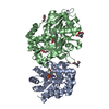
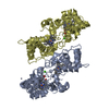
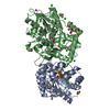
| Structure viewer | Molecule: MolmilJmol/JSmol |
|---|
- Downloads & links
-Download
| PDBx/mmCIF format | 1il0.cif.gz | 134.6 KB | Display | PDBx/mmCIF format |
|---|---|---|---|---|
| PDB format | pdb1il0.ent.gz | 104.3 KB | Display | PDB format |
| PDBx/mmJSON format | 1il0.json.gz | Tree view | PDBx/mmJSON format | |
| Others |  Other downloads Other downloads |
-Validation report
| Arichive directory | https://data.pdbj.org/pub/pdb/validation_reports/il/1il0ftp://data.pdbj.org/pub/pdb/validation_reports/il/1il0 | HTTPS FTP |
|---|
-Related structure data
| Related structure data | 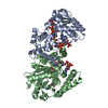 1f0yS S: Starting model for refinement |
|---|---|
| Similar structure data |


-Links
 PDBj
PDBj


- Assembly
| Deposited unit | 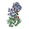
| ||||||||
|---|---|---|---|---|---|---|---|---|---|
| 1 |
| ||||||||
| Unit cell |
| ||||||||
| Details | The biological active dimer is contained within the asymetric unit |
-Components
| #1: Protein | / HCDH Mass: 32867.730 Da / Num. of mol.: 2 / Mutation: E170Q Source method: isolated from a genetically manipulated source Source: (gene. exp.) Homo sapiens (human) / Gene: HCDH / Plasmid: pET28 / Production host:  Escherichia coli (E. coli) Escherichia coli (E. coli)References: UniProt: Q16836, 3-hydroxyacyl-CoA dehydrogenase#2: Chemical | Acetoacetyl-CoA  Mass: 851.607 Da / Num. of mol.: 2 / Source method: obtained synthetically / Formula: C25H40N7O18P3S Mass: 851.607 Da / Num. of mol.: 2 / Source method: obtained synthetically / Formula: C25H40N7O18P3S#3: Chemical | Nicotinamide adenine dinucleotide  Mass: 663.425 Da / Num. of mol.: 2 / Source method: obtained synthetically / Formula: C21H27N7O14P2 / Comment: NAD*YM Mass: 663.425 Da / Num. of mol.: 2 / Source method: obtained synthetically / Formula: C21H27N7O14P2 / Comment: NAD*YM#4: Water | ChemComp-HOH / | Water Mass: 18.015 Da / Num. of mol.: 242 / Source method: isolated from a natural source / Formula: H2O Mass: 18.015 Da / Num. of mol.: 242 / Source method: isolated from a natural source / Formula: H2O |
|---|
-Experimental details
-Experiment
| Experiment | Method: X-RAY DIFFRACTION / Number of used crystals: 1 |
|---|
- Sample preparation
| Crystal | Density Matthews: 2.62 Å3/Da / Density % sol: 53.11 % | |||||||||||||||||||||||||
|---|---|---|---|---|---|---|---|---|---|---|---|---|---|---|---|---|---|---|---|---|---|---|---|---|---|---|
| Crystal grow | Temperature: 291 K / Method: vapor diffusion, hanging drop / pH: 6.5 Details: PEG 4000, N-[2-ACETAMIDO]-2-IMINODIACETIC ACID, pH 6.5, VAPOR DIFFUSION, HANGING DROP, temperature 291K | |||||||||||||||||||||||||
| Crystal grow | *PLUS Details: Barycki, J.J., (1999) Biochemistry, 38, 5786. | |||||||||||||||||||||||||
| Components of the solutions | *PLUS
|
-Data collection
| Diffraction | Mean temperature: 100 K |
|---|---|
| Diffraction source | Source: ROTATING ANODE / Type: RIGAKU RU200 / Wavelength: 1.5418 Å |
| Detector | Type: RIGAKU RAXIS IV / Detector: IMAGE PLATE / Date: Jan 18, 2000 / Details: Osmic MaxFlux |
| Radiation | Protocol: SINGLE WAVELENGTH / Monochromatic (M) / Laue (L): M / Scattering type: x-ray |
| Radiation wavelength | Wavelength: 1.5418 Å / Relative weight: 1 |
| Reflection | Resolution: 2.2→30 Å / Num. all: 36049 / Num. obs: 35973 / % possible obs: 99.8 % / Observed criterion σ(F): 0 / Observed criterion σ(I): 0 / Redundancy: 7.89 % / Biso Wilson estimate: 28.7 Å2 / Rmerge(I) obs: 0.09 / Net I/σ(I): 17.1 |
| Reflection shell | Resolution: 2.2→2.28 Å / Redundancy: 7.96 % / Rmerge(I) obs: 0.264 / Mean I/σ(I) obs: 9.4 / Num. unique all: 3530 / % possible all: 99.3 |
| Reflection | *PLUS Lowest resolution: 30 Å / Rmerge(I) obs: 0.09 |
| Reflection shell | *PLUS % possible obs: 99.3 % |
- Processing
| Software |
| ||||||||||||||||||||||||||||||||||||||||||||||||||||||||||||||||||||||||||||||||
|---|---|---|---|---|---|---|---|---|---|---|---|---|---|---|---|---|---|---|---|---|---|---|---|---|---|---|---|---|---|---|---|---|---|---|---|---|---|---|---|---|---|---|---|---|---|---|---|---|---|---|---|---|---|---|---|---|---|---|---|---|---|---|---|---|---|---|---|---|---|---|---|---|---|---|---|---|---|---|---|---|---|
| Refinement | Method to determine structure: MOLECULAR REPLACEMENT Starting model: PDB ENTRY 1F0Y Resolution: 2.2→30 Å / Rfactor Rfree error: 0.006 / Data cutoff high absF: 253077.14 / Data cutoff low absF: 0 / Isotropic thermal model: RESTRAINED / Cross valid method: THROUGHOUT / σ(F): 0 / σ(I): 0 / Stereochemistry target values: Engh & Huber
| ||||||||||||||||||||||||||||||||||||||||||||||||||||||||||||||||||||||||||||||||
| Displacement parameters | Biso mean: 29.9 Å2
| ||||||||||||||||||||||||||||||||||||||||||||||||||||||||||||||||||||||||||||||||
| Refine analyze |
| ||||||||||||||||||||||||||||||||||||||||||||||||||||||||||||||||||||||||||||||||
| Refinement step | Cycle: LAST / Resolution: 2.2→30 Å
| ||||||||||||||||||||||||||||||||||||||||||||||||||||||||||||||||||||||||||||||||
| Refine LS restraints |
| ||||||||||||||||||||||||||||||||||||||||||||||||||||||||||||||||||||||||||||||||
| LS refinement shell | Resolution: 2.2→2.34 Å / Rfactor Rfree error: 0.021 / Total num. of bins used: 6
| ||||||||||||||||||||||||||||||||||||||||||||||||||||||||||||||||||||||||||||||||
| Xplor file |
| ||||||||||||||||||||||||||||||||||||||||||||||||||||||||||||||||||||||||||||||||
| Software | *PLUS Name: CNS / Classification: refinement | ||||||||||||||||||||||||||||||||||||||||||||||||||||||||||||||||||||||||||||||||
| Refinement | *PLUS σ(F): 0 / % reflection Rfree: 5.1 % | ||||||||||||||||||||||||||||||||||||||||||||||||||||||||||||||||||||||||||||||||
| Solvent computation | *PLUS | ||||||||||||||||||||||||||||||||||||||||||||||||||||||||||||||||||||||||||||||||
| Displacement parameters | *PLUS Biso mean: 29.9 Å2 | ||||||||||||||||||||||||||||||||||||||||||||||||||||||||||||||||||||||||||||||||
| Refine LS restraints | *PLUS
| ||||||||||||||||||||||||||||||||||||||||||||||||||||||||||||||||||||||||||||||||
| LS refinement shell | *PLUS Rfactor Rfree: 0.34 / % reflection Rfree: 4.3 % / Rfactor Rwork: 0.282 / Rfactor obs: 0.282 |
