 Movie
Movie Controller

[English] 日本語
 Yorodumi
- PDB-1ib1: CRYSTAL STRUCTURE OF THE 14-3-3 ZETA:SEROTONIN N-ACETYLTRANSFERAS... -
+ Open data

- Basic information
| Entry | Database: PDB / ID: 1ib1 | ||||||
|---|---|---|---|---|---|---|---|
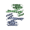
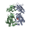
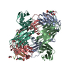
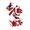
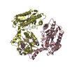
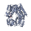
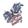
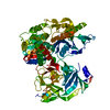
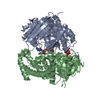
| Title | CRYSTAL STRUCTURE OF THE 14-3-3 ZETA:SEROTONIN N-ACETYLTRANSFERASE COMPLEX | ||||||
 Components Components |
| ||||||
 Keywords Keywords |  SIGNALING PROTEIN/TRANSFERASE / N-ACETYL TRANSFERASE / 14-3-3 / SIGNAL TRANSDUCTION / PROTEIN-PROTEIN COMPLEX / PHOSPHORYLATION / SIGNALING PROTEIN-TRANSFERASE COMPLEX SIGNALING PROTEIN/TRANSFERASE / N-ACETYL TRANSFERASE / 14-3-3 / SIGNAL TRANSDUCTION / PROTEIN-PROTEIN COMPLEX / PHOSPHORYLATION / SIGNALING PROTEIN-TRANSFERASE COMPLEX | ||||||
| Function / homology |  Function and homology informationaralkylamine N-acetyltransferase / aralkylamine N-acetyltransferase activity / melatonin biosynthetic process / N-terminal protein amino acid acetylation / Golgi reassembly / synaptic target recognition / regulation of synapse maturation / NOTCH4 Activation and Transmission of Signal to the Nucleus / respiratory system process / establishment of Golgi localization ...aralkylamine N-acetyltransferase / aralkylamine N-acetyltransferase activity / melatonin biosynthetic process / N-terminal protein amino acid acetylation / Golgi reassembly / synaptic target recognition / regulation of synapse maturation / NOTCH4 Activation and Transmission of Signal to the Nucleus / respiratory system process / establishment of Golgi localization / tube formation / Rap1 signalling / negative regulation of protein localization to nucleus / KSRP (KHSRP) binds and destabilizes mRNA / GP1b-IX-V activation signalling / Regulation of localization of FOXO transcription factors / Interleukin-3, Interleukin-5 and GM-CSF signaling / response to light stimulus / phosphoserine residue binding / Activation of BAD and translocation to mitochondria / protein targeting / SARS-CoV-2 targets host intracellular signalling and regulatory pathways / cellular response to glucose starvation / Chk1/Chk2(Cds1) mediated inactivation of Cyclin B:Cdk1 complex / SARS-CoV-1 targets host intracellular signalling and regulatory pathways / cellular response to cAMP / RHO GTPases activate PKNs / negative regulation of TORC1 signaling / ERK1 and ERK2 cascade / negative regulation of innate immune response / protein sequestering activity / hippocampal mossy fiber to CA3 synapse / regulation of ERK1 and ERK2 cascade / Translocation of SLC2A4 (GLUT4) to the plasma membrane / Deactivation of the beta-catenin transactivating complex / TP53 Regulates Metabolic Genes / Negative regulation of NOTCH4 signaling / lung development / regulation of protein stability / circadian rhythm / melanosome / DNA-binding transcription factor binding / angiogenesis / vesicle / transmembrane transporter binding / blood microparticle / cadherin binding / protein domain specific binding / protein phosphorylation / focal adhesion / glutamatergic synapse / ubiquitin protein ligase binding / negative regulation of apoptotic process / protein kinase binding / perinuclear region of cytoplasm / negative regulation of transcription by RNA polymerase II / signal transduction / extracellular space / RNA binding / extracellular exosome / nucleoplasm / identical protein binding / nucleus / cytosol / cytoplasm Function and homology informationaralkylamine N-acetyltransferase / aralkylamine N-acetyltransferase activity / melatonin biosynthetic process / N-terminal protein amino acid acetylation / Golgi reassembly / synaptic target recognition / regulation of synapse maturation / NOTCH4 Activation and Transmission of Signal to the Nucleus / respiratory system process / establishment of Golgi localization ...aralkylamine N-acetyltransferase / aralkylamine N-acetyltransferase activity / melatonin biosynthetic process / N-terminal protein amino acid acetylation / Golgi reassembly / synaptic target recognition / regulation of synapse maturation / NOTCH4 Activation and Transmission of Signal to the Nucleus / respiratory system process / establishment of Golgi localization / tube formation / Rap1 signalling / negative regulation of protein localization to nucleus / KSRP (KHSRP) binds and destabilizes mRNA / GP1b-IX-V activation signalling / Regulation of localization of FOXO transcription factors / Interleukin-3, Interleukin-5 and GM-CSF signaling / response to light stimulus / phosphoserine residue binding / Activation of BAD and translocation to mitochondria / protein targeting / SARS-CoV-2 targets host intracellular signalling and regulatory pathways / cellular response to glucose starvation / Chk1/Chk2(Cds1) mediated inactivation of Cyclin B:Cdk1 complex / SARS-CoV-1 targets host intracellular signalling and regulatory pathways / cellular response to cAMP / RHO GTPases activate PKNs / negative regulation of TORC1 signaling / ERK1 and ERK2 cascade / negative regulation of innate immune response / protein sequestering activity / hippocampal mossy fiber to CA3 synapse / regulation of ERK1 and ERK2 cascade / Translocation of SLC2A4 (GLUT4) to the plasma membrane / Deactivation of the beta-catenin transactivating complex / TP53 Regulates Metabolic Genes / Negative regulation of NOTCH4 signaling / lung development / regulation of protein stability / circadian rhythm / melanosome / DNA-binding transcription factor binding / angiogenesis / vesicle / transmembrane transporter binding / blood microparticle / cadherin binding / protein domain specific binding / protein phosphorylation / focal adhesion / glutamatergic synapse / ubiquitin protein ligase binding / negative regulation of apoptotic process / protein kinase binding / perinuclear region of cytoplasm / negative regulation of transcription by RNA polymerase II / signal transduction / extracellular space / RNA binding / extracellular exosome / nucleoplasm / identical protein binding / nucleus / cytosol / cytoplasmSimilarity search - Function | ||||||
| Biological species |  Homo sapiens (human) Homo sapiens (human) Ovis aries (sheep) Ovis aries (sheep) | ||||||
| Method | X-RAY DIFFRACTION / MOLECULAR REPLACEMENT / Resolution: 2.7 Å | ||||||
 Authors Authors | Obsil, T. / Ghirlando, R. / Klein, D.C. / Ganguly, S. / Dyda, F. | ||||||
 Citation Citation | Journal: Cell(Cambridge,Mass.) / Year: 2001 Title: Crystal structure of the 14-3-3zeta:serotonin N-acetyltransferase complex. a role for scaffolding in enzyme regulation. Authors: Obsil, T. / Ghirlando, R. / Klein, D.C. / Ganguly, S. / Dyda, F. | ||||||
| History |
| ||||||
| Remark 999 | SEQUENCE SEROTONIN N-ACETYLTRANSFERASE (CHAINS E,F,G,H) WAS EXPRESSED TRUNCATED (ONLY RESIDUES 1-201). |
- Structure visualization
| Structure viewer | Molecule: MolmilJmol/JSmol |
|---|
- Downloads & links
-Download
| PDBx/mmCIF format | 1ib1.cif.gz | 332.4 KB | Display | PDBx/mmCIF format |
|---|---|---|---|---|
| PDB format | pdb1ib1.ent.gz | 272.2 KB | Display | PDB format |
| PDBx/mmJSON format | 1ib1.json.gz | Tree view | PDBx/mmJSON format | |
| Others |  Other downloads Other downloads |
-Validation report
| Arichive directory | https://data.pdbj.org/pub/pdb/validation_reports/ib/1ib1ftp://data.pdbj.org/pub/pdb/validation_reports/ib/1ib1 | HTTPS FTP |
|---|
-Related structure data


-Links
 PDBj
PDBj


- Assembly
| Deposited unit | 
| ||||||||
|---|---|---|---|---|---|---|---|---|---|
| 1 | 
| ||||||||
| 2 | 
| ||||||||
| Unit cell |
|
-Components
| #1: Protein | Mass: 27777.092 Da / Num. of mol.: 4 Source method: isolated from a genetically manipulated source Source: (gene. exp.) Homo sapiens (human) / Gene: YWHAZ / Plasmid: PET14B / Species (production host): Escherichia coli / Production host:  Escherichia coli BL21(DE3) (bacteria) / Strain (production host): BL21(DE3) / References: UniProt: P63104 Escherichia coli BL21(DE3) (bacteria) / Strain (production host): BL21(DE3) / References: UniProt: P63104#2: Protein | Aralkylamine N-acetyltransferase / ARALKYLAMINE N-ACETYLTRANSFERASE / AA-NAT / SEROTONIN ACETYLASEMass: 22272.500 Da / Num. of mol.: 4 Source method: isolated from a genetically manipulated source Source: (gene. exp.) Ovis aries (sheep) / Gene: AANAT OR SNAT / Plasmid: PGEX-4T-1 / Production host: Escherichia coli (E. coli) / Strain (production host): BL21(DE3)PLYSReferences: UniProt: Q29495, aralkylamine N-acetyltransferase#3: Chemical | ChemComp-COT /   Mass: 967.771 Da / Num. of mol.: 4 / Source method: obtained synthetically / Formula: C33H48N9O17P3S Mass: 967.771 Da / Num. of mol.: 4 / Source method: obtained synthetically / Formula: C33H48N9O17P3S#4: Water | ChemComp-HOH / | Water Mass: 18.015 Da / Num. of mol.: 211 / Source method: isolated from a natural source / Formula: H2O Mass: 18.015 Da / Num. of mol.: 211 / Source method: isolated from a natural source / Formula: H2O |
|---|
-Experimental details
-Experiment
| Experiment | Method: X-RAY DIFFRACTION / Number of used crystals: 1 |
|---|
- Sample preparation
| Crystal | Density Matthews: 2.54 Å3/Da / Density % sol: 51.59 % | ||||||||||||||||||||||||||||||
|---|---|---|---|---|---|---|---|---|---|---|---|---|---|---|---|---|---|---|---|---|---|---|---|---|---|---|---|---|---|---|---|
| Crystal grow | Temperature: 277 K / Method: vapor diffusion, hanging drop / pH: 7.5 Details: PEG 4000, magnesium chloride, lithium sulfate, DTT, EDTA, pH 7.5, VAPOR DIFFUSION, HANGING DROP, temperature 277K | ||||||||||||||||||||||||||||||
| Crystal grow | *PLUS Temperature: 4 ℃ | ||||||||||||||||||||||||||||||
| Components of the solutions | *PLUS
|
-Data collection
| Diffraction | Mean temperature: 95 K |
|---|---|
| Diffraction source | Source: ROTATING ANODE / Type: RIGAKU RU200 / Wavelength: 1.5418 Å |
| Detector | Type: RIGAKU RAXIS IIC / Detector: IMAGE PLATE / Date: Apr 10, 2000 / Details: TOTAL REFLECTION MIRRORS |
| Radiation | Monochromator: NI FILTER / Protocol: SINGLE WAVELENGTH / Monochromatic (M) / Laue (L): M / Scattering type: x-ray |
| Radiation wavelength | Wavelength: 1.5418 Å / Relative weight: 1 |
| Reflection | Resolution: 2.7→20 Å / Num. all: 78001 / Num. obs: 44944 / % possible obs: 84.9 % / Observed criterion σ(I): 0 / Redundancy: 1.74 % / Biso Wilson estimate: 26 Å2 / Rmerge(I) obs: 0.102 / Rsym value: 0.102 / Net I/σ(I): 5.6 |
| Reflection shell | Resolution: 2.7→2.78 Å / Redundancy: 1.5 % / Rmerge(I) obs: 0.251 / Mean I/σ(I) obs: 2.09 / Num. unique all: 3188 / Rsym value: 0.251 / % possible all: 72 |
| Reflection | *PLUS Num. measured all: 78001 |
| Reflection shell | *PLUS % possible obs: 72 % |
- Processing
| Software |
| |||||||||||||||||||||||||
|---|---|---|---|---|---|---|---|---|---|---|---|---|---|---|---|---|---|---|---|---|---|---|---|---|---|---|
| Refinement | Method to determine structure: MOLECULAR REPLACEMENT Starting model: PDB ENTRY 1A4O AND 1CJW Resolution: 2.7→20 Å / Isotropic thermal model: isotropic / Cross valid method: THROUGHOUT / σ(F): 1 / σ(I): 0.5 / Stereochemistry target values: Engh & Huber
| |||||||||||||||||||||||||
| Displacement parameters | Biso mean: 16.3 Å2 | |||||||||||||||||||||||||
| Refinement step | Cycle: LAST / Resolution: 2.7→20 Å
| |||||||||||||||||||||||||
| Refine LS restraints |
| |||||||||||||||||||||||||
| LS refinement shell | Resolution: 2.7→2.82 Å / Rfactor Rfree error: 0.034
|
