 Movie
Movie Controller

[English] 日本語
 Yorodumi
- PDB-5z68: Structure of the recombination mediator protein RecF-ATP in RecFO... -
+ Open data

- Basic information
| Entry | Database: PDB / ID: 5z68 | ||||||
|---|---|---|---|---|---|---|---|
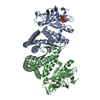
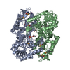
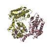
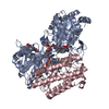
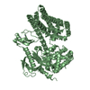
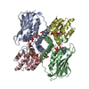
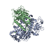
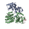
| Title | Structure of the recombination mediator protein RecF-ATP in RecFOR pathway | ||||||
 Components Components | DNA replication and repair protein RecF | ||||||
 Keywords Keywords |  DNA BINDING PROTEIN / recF / DNA repair / recombination mediator / RecFOR / Rad50 DNA BINDING PROTEIN / recF / DNA repair / recombination mediator / RecFOR / Rad50 | ||||||
| Function / homology |  Function and homology information Function and homology informationDNA synthesis involved in DNA repair / SOS response / double-strand break repair / single-stranded DNA binding / DNA replication / ATP binding / cytoplasmSimilarity search - Function | ||||||
| Biological species |  Caldanaerobacter subterraneus subsp. tengcongensis (bacteria) Caldanaerobacter subterraneus subsp. tengcongensis (bacteria) | ||||||
| Method | X-RAY DIFFRACTION / SYNCHROTRON / Resolution: 3 Å | ||||||
 Authors Authors | Tang, Q. / Liu, Y.-P. / Yan, X.-X. | ||||||
| Funding support |  China, 1items China, 1items
| ||||||
 Citation Citation | Journal: Sci Rep / Year: 2018 Title: ATP-dependent conformational change in ABC-ATPase RecF serves as a switch in DNA repair. Authors: Tang, Q. / Liu, Y.P. / Shan, H.H. / Tian, L.F. / Zhang, J.Z. / Yan, X.X. | ||||||
| History |
|
- Structure visualization
| Structure viewer | Molecule: MolmilJmol/JSmol |
|---|
- Downloads & links
-Download
| PDBx/mmCIF format | 5z68.cif.gz | 306 KB | Display | PDBx/mmCIF format |
|---|---|---|---|---|
| PDB format | pdb5z68.ent.gz | 253.7 KB | Display | PDB format |
| PDBx/mmJSON format | 5z68.json.gz | Tree view | PDBx/mmJSON format | |
| Others |  Other downloads Other downloads |
-Validation report
| Arichive directory | https://data.pdbj.org/pub/pdb/validation_reports/z6/5z68ftp://data.pdbj.org/pub/pdb/validation_reports/z6/5z68 | HTTPS FTP |
|---|
-Related structure data


-Links
 PDBj
PDBj


- Assembly
| Deposited unit | 
| ||||||||
|---|---|---|---|---|---|---|---|---|---|
| 1 | 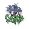
| ||||||||
| 2 | 
| ||||||||
| Unit cell |
|
-Components
| #1: Protein | Mass: 43389.980 Da / Num. of mol.: 4 Source method: isolated from a genetically manipulated source Source: (gene. exp.) Caldanaerobacter subterraneus subsp. tengcongensis (strain DSM 15242 / JCM 11007 / NBRC 100824 / MB4) (bacteria)Strain: DSM 15242 / JCM 11007 / NBRC 100824 / MB4 / Gene: recF, TTE0004 Production host: Escherichia coli 'BL21-Gold(DE3)pLysS AG' (bacteria)References: UniProt: Q8RDL3 #2: Chemical | ChemComp-ATP / Adenosine triphosphate  Mass: 507.181 Da / Num. of mol.: 4 / Source method: obtained synthetically / Formula: C10H16N5O13P3 / Comment: ATP, energy-carrying molecule*YM Mass: 507.181 Da / Num. of mol.: 4 / Source method: obtained synthetically / Formula: C10H16N5O13P3 / Comment: ATP, energy-carrying molecule*YM#3: Chemical | ChemComp-IMD / Imidazole  Mass: 69.085 Da / Num. of mol.: 21 / Source method: obtained synthetically / Formula: C3H5N2 Mass: 69.085 Da / Num. of mol.: 21 / Source method: obtained synthetically / Formula: C3H5N2#4: Chemical | ChemComp-MG /   Mass: 24.305 Da / Num. of mol.: 4 / Source method: obtained synthetically / Formula: Mg Mass: 24.305 Da / Num. of mol.: 4 / Source method: obtained synthetically / Formula: Mg#5: Water | ChemComp-HOH / | Water Mass: 18.015 Da / Num. of mol.: 104 / Source method: isolated from a natural source / Formula: H2O Mass: 18.015 Da / Num. of mol.: 104 / Source method: isolated from a natural source / Formula: H2O |
|---|
-Experimental details
-Experiment
| Experiment | Method: X-RAY DIFFRACTION / Number of used crystals: 1 |
|---|
- Sample preparation
| Crystal | Density Matthews: 3.92 Å3/Da / Density % sol: 68.6 % |
|---|---|
| Crystal grow | Temperature: 293 K / Method: vapor diffusion, hanging drop / pH: 8.5 / Details: 1.8M ammonium sulfate, 100 mM Tris-Hcl, pH 8.5 |
-Data collection
| Diffraction | Mean temperature: 100 K |
|---|---|
| Diffraction source | Source: SYNCHROTRON / Site: SSRF / Beamline: BL17U1 / Wavelength: 1.005 Å |
| Detector | Type: ADSC QUANTUM 315r / Detector: CCD / Date: Dec 19, 2014 |
| Radiation | Protocol: SINGLE WAVELENGTH / Monochromatic (M) / Laue (L): M / Scattering type: x-ray |
| Radiation wavelength | Wavelength: 1.005 Å / Relative weight: 1 |
| Reflection | Resolution: 3→20 Å / % possible obs: 99.9 % / Redundancy: 8.8 % / Net I/σ(I): 28.2 |
| Reflection shell | Resolution: 3→3.05 Å |
- Processing
| Software |
| ||||||||||||||||||||||||||||||||||||||||||||||||||||||||||||||||||||||||||||||||||||||||||||||||||||||||||||||||||||||||||||||||||||||||||||
|---|---|---|---|---|---|---|---|---|---|---|---|---|---|---|---|---|---|---|---|---|---|---|---|---|---|---|---|---|---|---|---|---|---|---|---|---|---|---|---|---|---|---|---|---|---|---|---|---|---|---|---|---|---|---|---|---|---|---|---|---|---|---|---|---|---|---|---|---|---|---|---|---|---|---|---|---|---|---|---|---|---|---|---|---|---|---|---|---|---|---|---|---|---|---|---|---|---|---|---|---|---|---|---|---|---|---|---|---|---|---|---|---|---|---|---|---|---|---|---|---|---|---|---|---|---|---|---|---|---|---|---|---|---|---|---|---|---|---|---|---|---|
| Refinement | Resolution: 3→19.863 Å / SU ML: 0.38 / Cross valid method: FREE R-VALUE / σ(F): 1.34 / Phase error: 30.26
| ||||||||||||||||||||||||||||||||||||||||||||||||||||||||||||||||||||||||||||||||||||||||||||||||||||||||||||||||||||||||||||||||||||||||||||
| Solvent computation | Shrinkage radii: 0.9 Å / VDW probe radii: 1.11 Å | ||||||||||||||||||||||||||||||||||||||||||||||||||||||||||||||||||||||||||||||||||||||||||||||||||||||||||||||||||||||||||||||||||||||||||||
| Refinement step | Cycle: LAST / Resolution: 3→19.863 Å
| ||||||||||||||||||||||||||||||||||||||||||||||||||||||||||||||||||||||||||||||||||||||||||||||||||||||||||||||||||||||||||||||||||||||||||||
| Refine LS restraints |
| ||||||||||||||||||||||||||||||||||||||||||||||||||||||||||||||||||||||||||||||||||||||||||||||||||||||||||||||||||||||||||||||||||||||||||||
| LS refinement shell |
|
