 Movie
Movie Controller

[English] 日本語
 Yorodumi
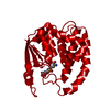
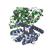
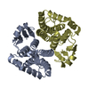
- PDB-1hnc: CRYSTAL STRUCTURE OF HUMAN CLASS MU GLUTATHIONE TRANSFERASE GSTM2... -
+ Open data

- Basic information
| Entry | Database: PDB / ID: 1hnc | ||||||
|---|---|---|---|---|---|---|---|
| Title | CRYSTAL STRUCTURE OF HUMAN CLASS MU GLUTATHIONE TRANSFERASE GSTM2-2: EFFECTS OF LATTICE PACKING ON CONFORMATIONAL HETEROGENEITY | ||||||
 Components Components | GLUTATHIONE S-TRANSFERASE | ||||||
 Keywords Keywords | TRANSFERASE(GLUTATHIONE) | ||||||
| Function / homology |  Function and homology information Function and homology informationnitrobenzene metabolic process / cellular detoxification of nitrogen compound / regulation of skeletal muscle contraction by regulation of release of sequestered calcium ion / hepoxilin biosynthetic process / glutathione binding / linoleic acid metabolic process / Glutathione conjugation / glutathione peroxidase activity / relaxation of cardiac muscle / intercellular bridge ...nitrobenzene metabolic process / cellular detoxification of nitrogen compound / regulation of skeletal muscle contraction by regulation of release of sequestered calcium ion / hepoxilin biosynthetic process / glutathione binding / linoleic acid metabolic process / Glutathione conjugation / glutathione peroxidase activity / relaxation of cardiac muscle / intercellular bridge / positive regulation of ryanodine-sensitive calcium-release channel activity / cellular response to caffeine / glutathione transferase / glutathione transferase activity / negative regulation of ryanodine-sensitive calcium-release channel activity / regulation of cardiac muscle contraction by regulation of the release of sequestered calcium ion / glutathione metabolic process / xenobiotic catabolic process / regulation of release of sequestered calcium ion into cytosol by sarcoplasmic reticulum / sarcoplasmic reticulum / fatty acid binding / signaling receptor binding / enzyme binding / protein homodimerization activity / extracellular exosome / cytosol / cytoplasmSimilarity search - Function | ||||||
| Biological species |  Homo sapiens (human) Homo sapiens (human) | ||||||
| Method | X-RAY DIFFRACTION / Resolution: 3 Å | ||||||
 Authors Authors | Raghunathan, S. / Chandross, R.J. / Kretsinger, R.H. / Allison, T.J. / Penington, C.J. / Rule, G.S. | ||||||
 Citation Citation | Journal: J.Mol.Biol. / Year: 1994 Title: Crystal structure of human class mu glutathione transferase GSTM2-2. Effects of lattice packing on conformational heterogeneity. Authors: Raghunathan, S. / Chandross, R.J. / Kretsinger, R.H. / Allison, T.J. / Penington, C.J. / Rule, G.S. #1: Journal: Biochemistry / Year: 1992Title: Mapping of the Substrate-Binding Site of a Human Class Mu Glutathione Transferase Using Nuclear Magnetic Resonance Spectroscopy Authors: Penington, C.R. / Rule, G.S. #2: Journal: Proc.Natl.Acad.Sci.USA / Year: 1991Title: Cloning, Expression, and Characterization of a Class Mu Glutathione Transferase from Human Muscle, the Product of the Gst4 Locus Authors: Vorachek, W.R. / Pearson, W.R. / Rule, G.S. | ||||||
| History |
|
- Structure visualization
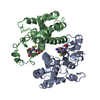
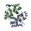
| Structure viewer | Molecule: MolmilJmol/JSmol |
|---|
- Downloads & links
-Download
| PDBx/mmCIF format | 1hnc.cif.gz | 183.7 KB | Display | PDBx/mmCIF format |
|---|---|---|---|---|
| PDB format | pdb1hnc.ent.gz | 148.9 KB | Display | PDB format |
| PDBx/mmJSON format | 1hnc.json.gz | Tree view | PDBx/mmJSON format | |
| Others |  Other downloads Other downloads |
-Validation report
| Arichive directory | https://data.pdbj.org/pub/pdb/validation_reports/hn/1hncftp://data.pdbj.org/pub/pdb/validation_reports/hn/1hnc | HTTPS FTP |
|---|
-Related structure data


-Links
 PDBj
PDBj

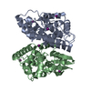
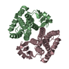
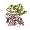
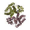
- Assembly
| Deposited unit | 
| ||||||||
|---|---|---|---|---|---|---|---|---|---|
| 1 | 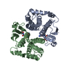
| ||||||||
| 2 | 
| ||||||||
| Unit cell |
| ||||||||
| Atom site foot note | 1: CIS PROLINE - PRO A 60 / 2: CIS PROLINE - PRO B 60 / 3: CIS PROLINE - PRO C 60 / 4: CIS PROLINE - PRO D 60 |
-Components
| #1: Protein | Mass: 25606.422 Da / Num. of mol.: 4 Source method: isolated from a genetically manipulated source Source: (gene. exp.) Homo sapiens (human) / References: UniProt: P28161, glutathione transferase#2: Chemical | ChemComp-GDN / 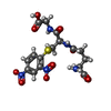  Mass: 473.415 Da / Num. of mol.: 4 / Source method: obtained synthetically / Formula: C16H19N5O10S Mass: 473.415 Da / Num. of mol.: 4 / Source method: obtained synthetically / Formula: C16H19N5O10SNonpolymer details | ALTHOUGH CRYSTALS WERE GROWN IN THE PRESENCE OF GLUTATHIONE S-(2,4 DINITROBENZENE), ONLY THE ...ALTHOUGH CRYSTALS WERE GROWN IN THE PRESENCE OF GLUTATHION | |
|---|
-Experimental details
-Experiment
| Experiment | Method: X-RAY DIFFRACTION |
|---|
- Sample preparation
| Crystal | Density Matthews: 2.44 Å3/Da / Density % sol: 49.49 % | ||||||||||||||||||||||||||||||||||||
|---|---|---|---|---|---|---|---|---|---|---|---|---|---|---|---|---|---|---|---|---|---|---|---|---|---|---|---|---|---|---|---|---|---|---|---|---|---|
| Crystal grow | *PLUS Temperature: 18 ℃ / Method: vapor diffusion, hanging drop / PH range low: 7.4 / PH range high: 6.8 | ||||||||||||||||||||||||||||||||||||
| Components of the solutions | *PLUS
|
-Data collection
| Radiation | Scattering type: x-ray |
|---|---|
| Radiation wavelength | Relative weight: 1 |
| Reflection | *PLUS Highest resolution: 2.8 Å / Num. obs: 24830 / % possible obs: 97 % / Redundancy: 5.1 % / Num. measured all: 127823 / Rmerge(I) obs: 0.102 / Biso Wilson estimate: 16.07 Å2 |
| Reflection shell | *PLUS Mean I/σ(I) obs: 5.2 |
- Processing
| Software |
| ||||||||||||||||||||||||||||||||||||||||||||||||||||||||||||
|---|---|---|---|---|---|---|---|---|---|---|---|---|---|---|---|---|---|---|---|---|---|---|---|---|---|---|---|---|---|---|---|---|---|---|---|---|---|---|---|---|---|---|---|---|---|---|---|---|---|---|---|---|---|---|---|---|---|---|---|---|---|
| Refinement | Resolution: 3→10 Å / σ(F): 0 Details: THE ELECTRON DENSITY FOR RESIDUES 203 - 217 IS POORLY DEFINED. THE TEMPERATURE FACTORS WERE NOT REFINED TO CONVERGENCE.
| ||||||||||||||||||||||||||||||||||||||||||||||||||||||||||||
| Refinement step | Cycle: LAST / Resolution: 3→10 Å
| ||||||||||||||||||||||||||||||||||||||||||||||||||||||||||||
| Refine LS restraints |
| ||||||||||||||||||||||||||||||||||||||||||||||||||||||||||||
| Software | *PLUS Name: X-PLOR / Classification: refinement | ||||||||||||||||||||||||||||||||||||||||||||||||||||||||||||
| Refinement | *PLUS Num. reflection all: 18827 / Rfactor obs: 0.25 | ||||||||||||||||||||||||||||||||||||||||||||||||||||||||||||
| Solvent computation | *PLUS | ||||||||||||||||||||||||||||||||||||||||||||||||||||||||||||
| Displacement parameters | *PLUS | ||||||||||||||||||||||||||||||||||||||||||||||||||||||||||||
| Refine LS restraints | *PLUS Type: x_angle_d / Dev ideal: 1.11 |
