 Movie
Movie Controller

[English] 日本語
 Yorodumi
- PDB-1xw5: Human glutathione s-transferase M2-2 (E.C.2.5.1.18)complexed with... -
+ Open data

- Basic information
| Entry | Database: PDB / ID: 1xw5 | ||||||
|---|---|---|---|---|---|---|---|
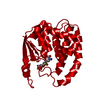
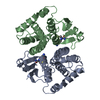
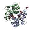
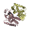
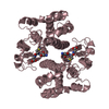
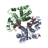
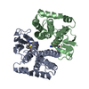
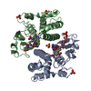
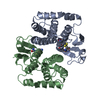
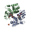
| Title | Human glutathione s-transferase M2-2 (E.C.2.5.1.18)complexed with glutathione, monoclinic crystal form | ||||||
 Components Components | Glutathione S-transferase Mu 2 | ||||||
 Keywords Keywords |  TRANSFERASE / GLUTATHIONE / CONJUGATION / DETOXIFICATION / CYTOSOLIC / DIMER / ACTIVE SITE TRANSFERASE / GLUTATHIONE / CONJUGATION / DETOXIFICATION / CYTOSOLIC / DIMER / ACTIVE SITE | ||||||
| Function / homology |  Function and homology information Function and homology informationnitrobenzene metabolic process / cellular detoxification of nitrogen compound / regulation of skeletal muscle contraction by regulation of release of sequestered calcium ion / hepoxilin biosynthetic process / glutathione binding / linoleic acid metabolic process / Glutathione conjugation / glutathione peroxidase activity / relaxation of cardiac muscle / intercellular bridge ...nitrobenzene metabolic process / cellular detoxification of nitrogen compound / regulation of skeletal muscle contraction by regulation of release of sequestered calcium ion / hepoxilin biosynthetic process / glutathione binding / linoleic acid metabolic process / Glutathione conjugation / glutathione peroxidase activity / relaxation of cardiac muscle / intercellular bridge / positive regulation of ryanodine-sensitive calcium-release channel activity / cellular response to caffeine / glutathione transferase / glutathione transferase activity / negative regulation of ryanodine-sensitive calcium-release channel activity / regulation of cardiac muscle contraction by regulation of the release of sequestered calcium ion / glutathione metabolic process / xenobiotic catabolic process / regulation of release of sequestered calcium ion into cytosol by sarcoplasmic reticulum / sarcoplasmic reticulum / fatty acid binding / signaling receptor binding / enzyme binding / protein homodimerization activity / extracellular exosome / cytosol / cytoplasmSimilarity search - Function | ||||||
| Biological species |  Homo sapiens (human) Homo sapiens (human) | ||||||
| Method | X-RAY DIFFRACTION / MOLECULAR REPLACEMENT / Resolution: 1.8 Å | ||||||
 Authors Authors | Patskovska, L.N. / Patskovsky, Y.V. / Almo, S.C. / Listowsky, I. | ||||||
 Citation Citation | Journal: To be Published Title: Structural Perturbation of the Active Site of Human Glutathione-S-Transferase M2-2 Upon Ligand Binding Authors: Patskovsky, Y. / Patskovska, L.N. / Almo, S.C. / Listowsky, I. #1: Journal: Acta Crystallogr.,Sect.D / Year: 1998Title: Expression, Crystallization and Preliminary X-Ray Analysis of Ligand-Free Human Glutathione S-Transferase M2-2 Authors: Patskovska, L.N. / Fedorov, A.A. / Patskovsky, Y.V. / Almo, S.C. / Listowsky, I. #2: Journal: J.Mol.Biol. / Year: 1994Title: Crystal Structure of Human Class Mu Glutathione Transferase Gstm2-2. Effects of Lattice Packing on Conformational Heterogeneity Authors: Raghunathan, S. / Chandross, R.J. / Kretsinger, R.H. / Allison, T.J. / Penington, C.J. / Rule, G.S. #3: Journal: Proc.Natl.Acad.Sci.USA / Year: 1991Title: Cloning, Expression, and Characterization of a Class-Mu Glutathione Transferase from Human Muscle, the Product of the Gst4 Locus Authors: Vorachek, W.R. / Pearson, W.R. / Rule, G.S. | ||||||
| History |
|
- Structure visualization
| Structure viewer | Molecule: MolmilJmol/JSmol |
|---|
- Downloads & links
-Download
| PDBx/mmCIF format | 1xw5.cif.gz | 112.6 KB | Display | PDBx/mmCIF format |
|---|---|---|---|---|
| PDB format | pdb1xw5.ent.gz | 86.3 KB | Display | PDB format |
| PDBx/mmJSON format | 1xw5.json.gz | Tree view | PDBx/mmJSON format | |
| Others |  Other downloads Other downloads |
-Validation report
| Arichive directory | https://data.pdbj.org/pub/pdb/validation_reports/xw/1xw5ftp://data.pdbj.org/pub/pdb/validation_reports/xw/1xw5 | HTTPS FTP |
|---|
-Related structure data
| Related structure data | 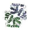 2gtuS S: Starting model for refinement |
|---|---|
| Similar structure data |


-Links
 PDBj
PDBj

- Assembly
| Deposited unit | 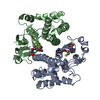
| ||||||||
|---|---|---|---|---|---|---|---|---|---|
| 1 |
| ||||||||
| Unit cell |
| ||||||||
| Details | Biological unit is a homodimer, the assymetric unit contains homodimer |
-Components
| #1: Protein | Mass: 25645.457 Da / Num. of mol.: 2 Source method: isolated from a genetically manipulated source Source: (gene. exp.) Homo sapiens (human) / Gene: GSTM2, GST4 / Plasmid: peT3a-hGSTM2 / Species (production host): Escherichia coli / Production host:  Escherichia coli BL21(DE3) (bacteria) / Strain (production host): BL21(DE3) / References: UniProt: P28161, glutathione transferase Escherichia coli BL21(DE3) (bacteria) / Strain (production host): BL21(DE3) / References: UniProt: P28161, glutathione transferase#2: Chemical | Glutathione  Mass: 307.323 Da / Num. of mol.: 2 / Source method: obtained synthetically / Formula: C10H17N3O6S Mass: 307.323 Da / Num. of mol.: 2 / Source method: obtained synthetically / Formula: C10H17N3O6S#3: Water | ChemComp-HOH / | Water Mass: 18.015 Da / Num. of mol.: 461 / Source method: isolated from a natural source / Formula: H2O Mass: 18.015 Da / Num. of mol.: 461 / Source method: isolated from a natural source / Formula: H2O |
|---|
-Experimental details
-Experiment
| Experiment | Method: X-RAY DIFFRACTION / Number of used crystals: 1 |
|---|
- Sample preparation
| Crystal | Density Matthews: 2.25 Å3/Da / Density % sol: 44.9 % |
|---|---|
| Crystal grow | Temperature: 290 K / Method: vapor diffusion / pH: 6.5 Details: 20% PEG4000, pH 6.50, VAPOR DIFFUSION, temperature 290K |
-Data collection
| Diffraction | Mean temperature: 100 K |
|---|---|
| Diffraction source | Source: ROTATING ANODE / Type: RIGAKU RU200 / Wavelength: 1.5418 / Wavelength: 1.5418 Å |
| Detector | Type: RIGAKU / Detector: IMAGE PLATE / Date: Oct 11, 2004 / Details: MIRRORS |
| Radiation | Monochromator: GRAPHITE CRYSTAL / Protocol: SINGLE WAVELENGTH / Monochromatic (M) / Laue (L): M / Scattering type: x-ray |
| Radiation wavelength | Wavelength: 1.5418 Å / Relative weight: 1 |
| Reflection | Resolution: 1.8→20 Å / Num. all: 43318 / Num. obs: 38664 / % possible obs: 89.3 % / Observed criterion σ(F): 0 / Observed criterion σ(I): 0 / Redundancy: 7.7 % / Biso Wilson estimate: 17.9 Å2 / Rmerge(I) obs: 0.064 / Rsym value: 0.062 / Net I/σ(I): 11.6 |
| Reflection shell | Resolution: 1.8→1.86 Å / Redundancy: 2.1 % / Rmerge(I) obs: 0.017 / Mean I/σ(I) obs: 2.2 / Num. unique all: 1864 / Rsym value: 0.019 / % possible all: 43.3 |
- Processing
| Software |
| ||||||||||||||||||||||||||||||||||||
|---|---|---|---|---|---|---|---|---|---|---|---|---|---|---|---|---|---|---|---|---|---|---|---|---|---|---|---|---|---|---|---|---|---|---|---|---|---|
| Refinement | Method to determine structure: MOLECULAR REPLACEMENT Starting model: PDB ENTRY 2GTU Resolution: 1.8→20 Å / Rfactor Rfree error: 0.007 / Data cutoff high absF: 10000 / Data cutoff low absF: 0.1 / Isotropic thermal model: RESTRAINED / Cross valid method: THROUGHOUT / σ(F): 1 / Stereochemistry target values: Engh & Huber
| ||||||||||||||||||||||||||||||||||||
| Solvent computation | Solvent model: FLAT MODEL / Bsol: 25.4053 Å2 / ksol: 0.318977 e/Å3 | ||||||||||||||||||||||||||||||||||||
| Displacement parameters | Biso mean: 23.9 Å2
| ||||||||||||||||||||||||||||||||||||
| Refine analyze |
| ||||||||||||||||||||||||||||||||||||
| Refinement step | Cycle: LAST / Resolution: 1.8→20 Å
| ||||||||||||||||||||||||||||||||||||
| Refine LS restraints |
| ||||||||||||||||||||||||||||||||||||
| LS refinement shell | Resolution: 1.8→1.91 Å / Rfactor Rfree error: 0.031 / Total num. of bins used: 6
| ||||||||||||||||||||||||||||||||||||
| Xplor file |
|
