 Movie
Movie Controller

[English] 日本語
 Yorodumi
- PDB-1h3g: Cyclomaltodextrinase from Flavobacterium sp. No. 92: from DNA seq... -
+ Open data

- Basic information
| Entry | Database: PDB / ID: 1h3g | ||||||
|---|---|---|---|---|---|---|---|
| Title | Cyclomaltodextrinase from Flavobacterium sp. No. 92: from DNA sequence to protein structure | ||||||
 Components Components | Cyclomaltodextrinase | ||||||
 Keywords Keywords | HYDROLASE / CYCLOMALTODEXTRINASE / GLYCOSIDASE | ||||||
| Function / homology |  Function and homology informationcyclomaltodextrinase / cyclomaltodextrinase activity / carbohydrate metabolic process / metal ion binding Function and homology informationcyclomaltodextrinase / cyclomaltodextrinase activity / carbohydrate metabolic process / metal ion bindingSimilarity search - Function | ||||||
| Biological species |  Flavobacterium sp. 92 (bacteria) Flavobacterium sp. 92 (bacteria) | ||||||
| Method | X-RAY DIFFRACTION / SYNCHROTRON / MAD / Resolution: 2.1 Å | ||||||
 Authors Authors | Fritzsche, H.B. / Schwede, T. / Jelakovic, S. / Schulz, G.E. | ||||||
 Citation Citation | Journal: Eur.J.Biochem. / Year: 2003 Title: Covalent and Three-Dimensional Structure of the Cyclodextrinase from Flavobacterium Sp. No. 92. Authors: Fritzsche, H.B. / Schwede, T. / Schulz, G.E. | ||||||
| History |
| ||||||
| Remark 650 | HELIX DETERMINATION METHOD: AUTHOR PROVIDED. |
- Structure visualization
| Structure viewer | Molecule: MolmilJmol/JSmol |
|---|
- Downloads & links
-Download
| PDBx/mmCIF format | 1h3g.cif.gz | 255.5 KB | Display | PDBx/mmCIF format |
|---|---|---|---|---|
| PDB format | pdb1h3g.ent.gz | 216.6 KB | Display | PDB format |
| PDBx/mmJSON format | 1h3g.json.gz | Tree view | PDBx/mmJSON format | |
| Others |  Other downloads Other downloads |
-Validation report
| Arichive directory | https://data.pdbj.org/pub/pdb/validation_reports/h3/1h3gftp://data.pdbj.org/pub/pdb/validation_reports/h3/1h3g | HTTPS FTP |
|---|
-Related structure data
| Similar structure data |
|---|


-Links
 PDBj
PDBj

- Assembly
| Deposited unit | 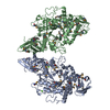
| ||||||||
|---|---|---|---|---|---|---|---|---|---|
| 1 | 
| ||||||||
| 2 | 
| ||||||||
| Unit cell |
| ||||||||
| Noncrystallographic symmetry (NCS) | NCS oper: (Code: given Matrix: (0.450916, 0.849463, -0.274019), Vector : |
-Components
| #1: Protein | Mass: 69259.969 Da / Num. of mol.: 2 Source method: isolated from a genetically manipulated source Source: (gene. exp.) Flavobacterium sp. 92 (bacteria) / Gene: cdase / Plasmid: PET22B+ / Production host: Escherichia coli BL21(DE3) (bacteria) / References: UniProt: Q8KKG0, cyclomaltodextrinase#2: Chemical | ChemComp-CA /   Mass: 40.078 Da / Num. of mol.: 4 / Source method: obtained synthetically / Formula: Ca Mass: 40.078 Da / Num. of mol.: 4 / Source method: obtained synthetically / Formula: Ca#3: Water | ChemComp-HOH / | Water Mass: 18.015 Da / Num. of mol.: 693 / Source method: isolated from a natural source / Formula: H2O Mass: 18.015 Da / Num. of mol.: 693 / Source method: isolated from a natural source / Formula: H2OCompound details | DEGRADATION OF CYCLODEXTRINS AND LINEAR MALTOOLIGOSACCHARIDES, HYDROLIZATION OF DIFFERENT ...DEGRADATIO | |
|---|
-Experimental details
-Experiment
| Experiment | Method: X-RAY DIFFRACTION / Number of used crystals: 1 |
|---|
- Sample preparation
| Crystal | Density Matthews: 2.74 Å3/Da / Density % sol: 55 % | ||||||||||||||||||||||||||||
|---|---|---|---|---|---|---|---|---|---|---|---|---|---|---|---|---|---|---|---|---|---|---|---|---|---|---|---|---|---|
| Crystal grow | pH: 7.5 / Details: pH 7.50 | ||||||||||||||||||||||||||||
| Crystal grow | *PLUS pH: 7.5 / Method: vapor diffusion, hanging drop | ||||||||||||||||||||||||||||
| Components of the solutions | *PLUS
|
-Data collection
| Diffraction | Mean temperature: 100 K |
|---|---|
| Diffraction source | Source: SYNCHROTRON / Site: EMBL/DESY, HAMBURG  / Beamline: BW7A / Wavelength: 0.9393 / Beamline: BW7A / Wavelength: 0.9393 |
| Detector | Type: MARRESEARCH / Detector: CCD / Date: Nov 3, 2001 / Details: MIRRORS |
| Radiation | Monochromator: DOUBLE CRYSTAL FOCUSSING / Protocol: MAD / Monochromatic (M) / Laue (L): M / Scattering type: x-ray |
| Radiation wavelength | Wavelength: 0.9393 Å / Relative weight: 1 |
| Reflection | Resolution: 2.1→21 Å / Num. obs: 86572 / % possible obs: 99.9 % / Observed criterion σ(I): 1.8 / Redundancy: 7.3 % / Rmerge(I) obs: 0.06 / Net I/σ(I): 10.4 |
| Reflection shell | Resolution: 2.1→2.18 Å / Redundancy: 7.3 % / Rmerge(I) obs: 0.398 / Mean I/σ(I) obs: 1.9 / % possible all: 99.9 |
| Reflection | *PLUS Highest resolution: 2.1 Å / Lowest resolution: 25 Å / Redundancy: 7.3 % / Num. measured all: 629678 / Rmerge(I) obs: 0.06 |
| Reflection shell | *PLUS Highest resolution: 2.08 Å / % possible obs: 99.9 % / Redundancy: 7.3 % / Num. unique obs: 8625 / Rmerge(I) obs: 0.4 / Mean I/σ(I) obs: 1.9 |
- Processing
| Software |
| ||||||||||||||||||||
|---|---|---|---|---|---|---|---|---|---|---|---|---|---|---|---|---|---|---|---|---|---|
| Refinement | Method to determine structure: MAD / Resolution: 2.1→21.1 Å / SU B: 6.4 / SU ML: 0.17 / Cross valid method: THROUGHOUT / ESU R: 0.21 / ESU R Free: 0.17 Details: FIRST CNS WAS USED FOR REFINEMENT FOLLOWED BY TLS REFINEMENT IN REFMAC
| ||||||||||||||||||||
| Displacement parameters | Biso mean: 27.8 Å2
| ||||||||||||||||||||
| Refinement step | Cycle: LAST / Resolution: 2.1→21.1 Å
| ||||||||||||||||||||
| Refinement | *PLUS Lowest resolution: 21 Å / Num. reflection obs: 78164 | ||||||||||||||||||||
| Solvent computation | *PLUS | ||||||||||||||||||||
| Displacement parameters | *PLUS | ||||||||||||||||||||
| Refine LS restraints | *PLUS
| ||||||||||||||||||||
| LS refinement shell | *PLUS Highest resolution: 2.08 Å / Lowest resolution: 2.13 Å / Rfactor Rfree: 0.278 / Rfactor Rwork: 0.219 / Num. reflection obs: 4817 |
