 Movie
Movie Controller


+ Open data

- Basic information
| Entry | Database: PDB / ID: 5u2a | ||||||
|---|---|---|---|---|---|---|---|
| Title | Crystal structure of Brucella canis Acyl-CoA Synthetase | ||||||
 Components Components | AMP-dependent synthetase and ligase | ||||||
 Keywords Keywords |  LIGASE / SSGCID / NIH / NIAID / SBRI / UW / BERYLLIUM / SYNTHETASE / ACYL-COA / Structural Genomics / Seattle Structural Genomics Center for Infectious Disease LIGASE / SSGCID / NIH / NIAID / SBRI / UW / BERYLLIUM / SYNTHETASE / ACYL-COA / Structural Genomics / Seattle Structural Genomics Center for Infectious Disease | ||||||
| Function / homology |  Function and homology information Function and homology information | ||||||
| Biological species | Brucella canis | ||||||
| Method | X-RAY DIFFRACTION / SYNCHROTRON / MOLECULAR REPLACEMENT / Resolution: 2.5 Å | ||||||
 Authors Authors | Seattle Structural Genomics Center for Infectious Disease (SSGCID) / Fox III, D. / Abendroth, J. | ||||||
 Citation Citation | Journal: To Be Published Title: Crystal structure of Brucella canis Acyl-CoA Synthetase Authors: Seattle Structural Genomics Center for Infectious Disease (SSGCID) / Fox III, D. / Abendroth, J. | ||||||
| History |
|
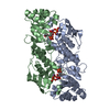
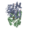
- Structure visualization
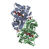
| Structure viewer | Molecule: MolmilJmol/JSmol |
|---|
- Downloads & links
-Download
| PDBx/mmCIF format | 5u2a.cif.gz | 216.9 KB | Display | PDBx/mmCIF format |
|---|---|---|---|---|
| PDB format | pdb5u2a.ent.gz | 172 KB | Display | PDB format |
| PDBx/mmJSON format | 5u2a.json.gz | Tree view | PDBx/mmJSON format | |
| Others |  Other downloads Other downloads |
-Validation report
| Arichive directory | https://data.pdbj.org/pub/pdb/validation_reports/u2/5u2aftp://data.pdbj.org/pub/pdb/validation_reports/u2/5u2a | HTTPS FTP |
|---|
-Related structure data
| Related structure data | 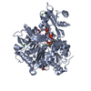 1pg4S S: Starting model for refinement |
|---|---|
| Similar structure data | |
| Other databases |


-Links
 PDBj
PDBj
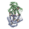
- Assembly
| Deposited unit | 
| ||||||||
|---|---|---|---|---|---|---|---|---|---|
| 1 |
| ||||||||
| Unit cell |
|
-Components
| #1: Protein | Mass: 60664.629 Da / Num. of mol.: 1 Source method: isolated from a genetically manipulated source Source: (gene. exp.)  Brucella canis (strain ATCC 23365 / NCTC 10854) (bacteria) Brucella canis (strain ATCC 23365 / NCTC 10854) (bacteria)Strain: ATCC 23365 / NCTC 10854 / Gene: BCAN_B0452 / Plasmid: BG1861 / Production host: ESCHERICHIA COLI (E. coli) / Strain (production host): BL21(DE3) / References: UniProt: A9MB96 |
|---|---|
| #2: Chemical | ChemComp-CL / Chloride  Mass: 35.453 Da / Num. of mol.: 1 / Source method: obtained synthetically / Formula: Cl Mass: 35.453 Da / Num. of mol.: 1 / Source method: obtained synthetically / Formula: Cl |
| #3: Water | ChemComp-HOH / Water Mass: 18.015 Da / Num. of mol.: 41 / Source method: isolated from a natural source / Formula: H2O Mass: 18.015 Da / Num. of mol.: 41 / Source method: isolated from a natural source / Formula: H2O |
-Experimental details
-Experiment
| Experiment | Method: X-RAY DIFFRACTION / Number of used crystals: 1 |
|---|
- Sample preparation
| Crystal | Density Matthews: 3.45 Å3/Da / Density % sol: 64.32 % |
|---|---|
| Crystal grow | Temperature: 289 K / Method: vapor diffusion, sitting drop / pH: 8.5 Details: ACETYL COA SYNTHETASE FROM BRUCELLA CANIS ATCC 23365 (BRCAA.00629.A.B1.PW37891) AT 26MG/ML (25 MM TRIS PH 8.0, 200 MM NACL, 1% GLYCEROL, 1 MM TCEP) WAS SET UP IN SPARSE CRYSTALLIZATION ...Details: ACETYL COA SYNTHETASE FROM BRUCELLA CANIS ATCC 23365 (BRCAA.00629.A.B1.PW37891) AT 26MG/ML (25 MM TRIS PH 8.0, 200 MM NACL, 1% GLYCEROL, 1 MM TCEP) WAS SET UP IN SPARSE CRYSTALLIZATION TRIALS AT 16C. CRYSTALS WERE PRODUCED BY SITTING DROP VAPOR DIFFUSION WITH AN EQUAL VOLUME OF PROTEIN AND SPARSE SCREEN MORPHEUS CONDITION A9 (0.1M TRIS(BASE), 0.1M BICINE, PH8.5, 0.06M MAGNESIUM CHLORIDE HEXAHYDRATE, 0.06M CALCIUM CHLORIDE DIHYDRATE, 20% PEG500 MME, 10% PEG20,000) AND DIRECTLY CRYO-PROTECTED. CRYSTAL ID 273132A9,RDJ0-10, CLSI-08ID, VAPOR DIFFUSION, SITTING DROP, TEMPERATURE 289K PH range: 8.5 |
-Data collection
| Diffraction | Mean temperature: 100 K |
|---|---|
| Diffraction source | Source: SYNCHROTRON / Site: CLSI  / Beamline: 08ID-1 / Wavelength: 0.97949 Å / Beamline: 08ID-1 / Wavelength: 0.97949 Å |
| Detector | Type: RAYONIX MX-225 / Detector: CCD / Date: May 27, 2016 |
| Radiation | Protocol: SINGLE WAVELENGTH / Monochromatic (M) / Laue (L): M / Scattering type: x-ray |
| Radiation wavelength | Wavelength: 0.97949 Å / Relative weight: 1 |
| Reflection | Resolution: 2.5→50 Å / Num. obs: 28526 / % possible obs: 99.6 % / Observed criterion σ(I): -3 / Redundancy: 4.8 % / Biso Wilson estimate: 70.73 Å2 / CC1/2: 0.999 / Rmerge(I) obs: 0.047 / Net I/σ(I): 20.27 |
| Reflection shell | Resolution: 2.5→2.56 Å / Redundancy: 5 % / Rmerge(I) obs: 0.512 / Mean I/σ(I) obs: 3.36 / Num. unique all: 10440 / CC1/2: 0.944 / % possible all: 99.9 |
- Processing
| Software |
| |||||||||||||||||||||||||||||||||||||||||||||||||||||||||||||||||||||||||||||||||||||||||||||||||||||||||||||||||||||||||||||||||||||||||||||||||||||||||||||||||||||||||||||||
|---|---|---|---|---|---|---|---|---|---|---|---|---|---|---|---|---|---|---|---|---|---|---|---|---|---|---|---|---|---|---|---|---|---|---|---|---|---|---|---|---|---|---|---|---|---|---|---|---|---|---|---|---|---|---|---|---|---|---|---|---|---|---|---|---|---|---|---|---|---|---|---|---|---|---|---|---|---|---|---|---|---|---|---|---|---|---|---|---|---|---|---|---|---|---|---|---|---|---|---|---|---|---|---|---|---|---|---|---|---|---|---|---|---|---|---|---|---|---|---|---|---|---|---|---|---|---|---|---|---|---|---|---|---|---|---|---|---|---|---|---|---|---|---|---|---|---|---|---|---|---|---|---|---|---|---|---|---|---|---|---|---|---|---|---|---|---|---|---|---|---|---|---|---|---|---|---|
| Refinement | Method to determine structure: MOLECULAR REPLACEMENT Starting model: 1pg4 Resolution: 2.5→41.918 Å / SU ML: 0.31 / Cross valid method: FREE R-VALUE / σ(F): 1.34 / Phase error: 28.95
| |||||||||||||||||||||||||||||||||||||||||||||||||||||||||||||||||||||||||||||||||||||||||||||||||||||||||||||||||||||||||||||||||||||||||||||||||||||||||||||||||||||||||||||||
| Solvent computation | Shrinkage radii: 0.9 Å / VDW probe radii: 1.11 Å | |||||||||||||||||||||||||||||||||||||||||||||||||||||||||||||||||||||||||||||||||||||||||||||||||||||||||||||||||||||||||||||||||||||||||||||||||||||||||||||||||||||||||||||||
| Displacement parameters | Biso mean: 70.73 Å2 | |||||||||||||||||||||||||||||||||||||||||||||||||||||||||||||||||||||||||||||||||||||||||||||||||||||||||||||||||||||||||||||||||||||||||||||||||||||||||||||||||||||||||||||||
| Refinement step | Cycle: LAST / Resolution: 2.5→41.918 Å
| |||||||||||||||||||||||||||||||||||||||||||||||||||||||||||||||||||||||||||||||||||||||||||||||||||||||||||||||||||||||||||||||||||||||||||||||||||||||||||||||||||||||||||||||
| Refine LS restraints |
| |||||||||||||||||||||||||||||||||||||||||||||||||||||||||||||||||||||||||||||||||||||||||||||||||||||||||||||||||||||||||||||||||||||||||||||||||||||||||||||||||||||||||||||||
| LS refinement shell |
| |||||||||||||||||||||||||||||||||||||||||||||||||||||||||||||||||||||||||||||||||||||||||||||||||||||||||||||||||||||||||||||||||||||||||||||||||||||||||||||||||||||||||||||||
| Refinement TLS params. | Method: refined / Refine-ID: X-RAY DIFFRACTION
| |||||||||||||||||||||||||||||||||||||||||||||||||||||||||||||||||||||||||||||||||||||||||||||||||||||||||||||||||||||||||||||||||||||||||||||||||||||||||||||||||||||||||||||||
| Refinement TLS group |
|
