 Movie
Movie Controller

[English] 日本語
 Yorodumi
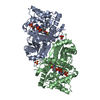
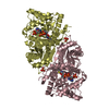
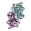
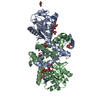
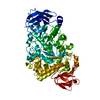
- PDB-5hid: BRAF Kinase domain b3aC loop deletion mutant in complex with AZ628 -
+ Open data

- Basic information
| Entry | Database: PDB / ID: 5hid | ||||||
|---|---|---|---|---|---|---|---|
| Title | BRAF Kinase domain b3aC loop deletion mutant in complex with AZ628 | ||||||
 Components Components | Serine/threonine-protein kinase B-raf | ||||||
 Keywords Keywords | TRANSFERASE/TRANSFERASE INHIBITOR / TRANSFERASE-TRANSFERASE INHIBITOR complex | ||||||
| Function / homology |  Function and homology information Function and homology informationtrehalose metabolism in response to stress / CD4-positive, alpha-beta T cell differentiation / CD4-positive or CD8-positive, alpha-beta T cell lineage commitment / negative regulation of synaptic vesicle exocytosis / head morphogenesis / Signalling to p38 via RIT and RIN / myeloid progenitor cell differentiation / ARMS-mediated activation / SHOC2 M1731 mutant abolishes MRAS complex function / Gain-of-function MRAS complexes activate RAF signaling ...trehalose metabolism in response to stress / CD4-positive, alpha-beta T cell differentiation / CD4-positive or CD8-positive, alpha-beta T cell lineage commitment / negative regulation of synaptic vesicle exocytosis / head morphogenesis / Signalling to p38 via RIT and RIN / myeloid progenitor cell differentiation / ARMS-mediated activation / SHOC2 M1731 mutant abolishes MRAS complex function / Gain-of-function MRAS complexes activate RAF signaling / endothelial cell apoptotic process / negative regulation of fibroblast migration / positive regulation of glucose transmembrane transport / establishment of protein localization to membrane / mitogen-activated protein kinase kinase binding / regulation of T cell differentiation / Negative feedback regulation of MAPK pathway / positive regulation of axonogenesis / Frs2-mediated activation /  stress fiber assembly / positive regulation of axon regeneration / face development / synaptic vesicle exocytosis / somatic stem cell population maintenance / MAP kinase kinase activity / thyroid gland development / MAP kinase kinase kinase activity / negative regulation of endothelial cell apoptotic process / positive regulation of substrate adhesion-dependent cell spreading / positive regulation of stress fiber assembly / response to cAMP / cellular response to calcium ion / ERK1 and ERK2 cascade / substrate adhesion-dependent cell spreading / cellular response to nerve growth factor stimulus / thymus development / long-term synaptic potentiation / animal organ morphogenesis / Spry regulation of FGF signaling / RAF activation / Signaling by high-kinase activity BRAF mutants / visual learning / MAP2K and MAPK activation / epidermal growth factor receptor signaling pathway / response to peptide hormone / Negative regulation of MAPK pathway / Signaling by RAF1 mutants / Signaling by moderate kinase activity BRAF mutants / Paradoxical activation of RAF signaling by kinase inactive BRAF / Signaling downstream of RAS mutants / MAPK cascade / Signaling by BRAF and RAF1 fusions / cellular response to xenobiotic stimulus / presynapse / positive regulation of peptidyl-serine phosphorylation / T cell differentiation in thymus / T cell receptor signaling pathway / regulation of cell population proliferation / cell body / scaffold protein binding / negative regulation of neuron apoptotic process / Ras protein signal transduction / positive regulation of ERK1 and ERK2 cascade / non-specific serine/threonine protein kinase / protein kinase activity / neuron projection / protein phosphorylation / intracellular membrane-bounded organelle / protein serine kinase activity / protein serine/threonine kinase activity / calcium ion binding / protein-containing complex binding / positive regulation of gene expression / negative regulation of apoptotic process / mitochondrion / ATP binding / identical protein binding / nucleus / plasma membrane / cytosol stress fiber assembly / positive regulation of axon regeneration / face development / synaptic vesicle exocytosis / somatic stem cell population maintenance / MAP kinase kinase activity / thyroid gland development / MAP kinase kinase kinase activity / negative regulation of endothelial cell apoptotic process / positive regulation of substrate adhesion-dependent cell spreading / positive regulation of stress fiber assembly / response to cAMP / cellular response to calcium ion / ERK1 and ERK2 cascade / substrate adhesion-dependent cell spreading / cellular response to nerve growth factor stimulus / thymus development / long-term synaptic potentiation / animal organ morphogenesis / Spry regulation of FGF signaling / RAF activation / Signaling by high-kinase activity BRAF mutants / visual learning / MAP2K and MAPK activation / epidermal growth factor receptor signaling pathway / response to peptide hormone / Negative regulation of MAPK pathway / Signaling by RAF1 mutants / Signaling by moderate kinase activity BRAF mutants / Paradoxical activation of RAF signaling by kinase inactive BRAF / Signaling downstream of RAS mutants / MAPK cascade / Signaling by BRAF and RAF1 fusions / cellular response to xenobiotic stimulus / presynapse / positive regulation of peptidyl-serine phosphorylation / T cell differentiation in thymus / T cell receptor signaling pathway / regulation of cell population proliferation / cell body / scaffold protein binding / negative regulation of neuron apoptotic process / Ras protein signal transduction / positive regulation of ERK1 and ERK2 cascade / non-specific serine/threonine protein kinase / protein kinase activity / neuron projection / protein phosphorylation / intracellular membrane-bounded organelle / protein serine kinase activity / protein serine/threonine kinase activity / calcium ion binding / protein-containing complex binding / positive regulation of gene expression / negative regulation of apoptotic process / mitochondrion / ATP binding / identical protein binding / nucleus / plasma membrane / cytosolSimilarity search - Function | ||||||
| Biological species |  Homo sapiens (human) Homo sapiens (human) | ||||||
| Method | X-RAY DIFFRACTION / SYNCHROTRON / MOLECULAR REPLACEMENT / Resolution: 2.5 Å | ||||||
 Authors Authors | Whalen, D.M. / Foster, S.A. / Ozen, A. / Wongchenko, M. / Yin, J. / Schaefer, G. / Mayfield, J. / Chmielecki, J. / Stephens, P. / Albacker, L. ...Whalen, D.M. / Foster, S.A. / Ozen, A. / Wongchenko, M. / Yin, J. / Schaefer, G. / Mayfield, J. / Chmielecki, J. / Stephens, P. / Albacker, L. / Yan, Y. / Song, K. / Hatzivassiliou, G. / Eigenbrot, C. / Yu, C. / Shaw, A.S. / Manning, G. / Skelton, N.J. / Hymowitz, S.G. / Malek, S. | ||||||
 Citation Citation | Journal: Cancer Cell / Year: 2016 Title: Activation Mechanism of Oncogenic Deletion Mutations in BRAF, EGFR, and HER2. Authors: Foster, S.A. / Whalen, D.M. / Ozen, A. / Wongchenko, M.J. / Yin, J. / Yen, I. / Schaefer, G. / Mayfield, J.D. / Chmielecki, J. / Stephens, P.J. / Albacker, L.A. / Yan, Y. / Song, K. / ...Authors: Foster, S.A. / Whalen, D.M. / Ozen, A. / Wongchenko, M.J. / Yin, J. / Yen, I. / Schaefer, G. / Mayfield, J.D. / Chmielecki, J. / Stephens, P.J. / Albacker, L.A. / Yan, Y. / Song, K. / Hatzivassiliou, G. / Eigenbrot, C. / Yu, C. / Shaw, A.S. / Manning, G. / Skelton, N.J. / Hymowitz, S.G. / Malek, S. | ||||||
| History |
|
- Structure visualization
| Structure viewer | Molecule: MolmilJmol/JSmol |
|---|
- Downloads & links
-Download
| PDBx/mmCIF format | 5hid.cif.gz | 226.5 KB | Display | PDBx/mmCIF format |
|---|---|---|---|---|
| PDB format | pdb5hid.ent.gz | 181.8 KB | Display | PDB format |
| PDBx/mmJSON format | 5hid.json.gz | Tree view | PDBx/mmJSON format | |
| Others |  Other downloads Other downloads |
-Validation report
| Arichive directory | https://data.pdbj.org/pub/pdb/validation_reports/hi/5hidftp://data.pdbj.org/pub/pdb/validation_reports/hi/5hid | HTTPS FTP |
|---|
-Related structure data
| Related structure data | 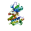 5hi2SC  5hibC 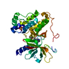 5hicC 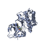 5hieC S: Starting model for refinement C: citing same article ( |
|---|---|
| Similar structure data |


-Links
 PDBj
PDBj


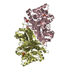
- Assembly
| Deposited unit | 
| ||||||||
|---|---|---|---|---|---|---|---|---|---|
| 1 |
| ||||||||
| Unit cell |
|
-Components
| #1: Protein | Mass: 32359.061 Da / Num. of mol.: 2 Source method: isolated from a genetically manipulated source Source: (gene. exp.) Homo sapiens (human) / Gene: BRAF, BRAF1, RAFB1 / Production host:  Escherichia coli (E. coli) Escherichia coli (E. coli)References: UniProt: P15056, non-specific serine/threonine protein kinase#2: Chemical |   Mass: 451.520 Da / Num. of mol.: 2 / Source method: obtained synthetically / Formula: C27H25N5O2 Mass: 451.520 Da / Num. of mol.: 2 / Source method: obtained synthetically / Formula: C27H25N5O2#3: Chemical | Diethylene glycol  Mass: 106.120 Da / Num. of mol.: 2 / Source method: isolated from a natural source / Formula: C4H10O3 Mass: 106.120 Da / Num. of mol.: 2 / Source method: isolated from a natural source / Formula: C4H10O3#4: Water | ChemComp-HOH / | Water Mass: 18.015 Da / Num. of mol.: 113 / Source method: isolated from a natural source / Formula: H2O Mass: 18.015 Da / Num. of mol.: 113 / Source method: isolated from a natural source / Formula: H2O |
|---|
-Experimental details
-Experiment
| Experiment | Method: X-RAY DIFFRACTION / Number of used crystals: 1 |
|---|
- Sample preparation
| Crystal | Density Matthews: 2.17 Å3/Da / Density % sol: 43.23 % |
|---|---|
| Crystal grow | Temperature: 292 K / Method: vapor diffusion, hanging drop / Details: 20% PEG 3350, and 0.2M Potassium Nitrate |
-Data collection
| Diffraction | Mean temperature: 110 K |
|---|---|
| Diffraction source | Source: SYNCHROTRON / Site: SSRL  / Beamline: BL12-2 / Wavelength: 0.9795 Å / Beamline: BL12-2 / Wavelength: 0.9795 Å |
| Detector | Type: MARMOSAIC 325 mm CCD / Detector: CCD / Date: Jul 22, 2015 |
| Radiation | Monochromator: Liquid nitrogen-cooled double crystal, non fixed exit slit. Si(111) Protocol: SINGLE WAVELENGTH / Monochromatic (M) / Laue (L): M / Scattering type: x-ray |
| Radiation wavelength | Wavelength: 0.9795 Å / Relative weight: 1 |
| Reflection | Resolution: 2.5→47.86 Å / Biso Wilson estimate: 44.14 Å2 |
| Reflection shell | Resolution: 2.5→2.65 Å / Mean I/σ(I) obs: 1.52 / % possible all: 71.3 |
- Processing
| Software |
| |||||||||||||||||||||||||||||||||||||||||||||||||||||||||||||||||||||||||||||||||||||||||||||||||||||||||||||||||||||||||||||
|---|---|---|---|---|---|---|---|---|---|---|---|---|---|---|---|---|---|---|---|---|---|---|---|---|---|---|---|---|---|---|---|---|---|---|---|---|---|---|---|---|---|---|---|---|---|---|---|---|---|---|---|---|---|---|---|---|---|---|---|---|---|---|---|---|---|---|---|---|---|---|---|---|---|---|---|---|---|---|---|---|---|---|---|---|---|---|---|---|---|---|---|---|---|---|---|---|---|---|---|---|---|---|---|---|---|---|---|---|---|---|---|---|---|---|---|---|---|---|---|---|---|---|---|---|---|---|
| Refinement | Method to determine structure: MOLECULAR REPLACEMENT Starting model: 5HI2 (partially refined) Resolution: 2.5→47.86 Å / Cor.coef. Fo:Fc: 0.934 / Cor.coef. Fo:Fc free: 0.9204 / SU R Cruickshank DPI: 1.029 / Cross valid method: THROUGHOUT / σ(F): 0 / SU R Blow DPI: 1.296 / SU Rfree Blow DPI: 0.336 / SU Rfree Cruickshank DPI: 0.336
| |||||||||||||||||||||||||||||||||||||||||||||||||||||||||||||||||||||||||||||||||||||||||||||||||||||||||||||||||||||||||||||
| Displacement parameters | Biso mean: 41.46 Å2
| |||||||||||||||||||||||||||||||||||||||||||||||||||||||||||||||||||||||||||||||||||||||||||||||||||||||||||||||||||||||||||||
| Refine analyze | Luzzati coordinate error obs: 0.364 Å | |||||||||||||||||||||||||||||||||||||||||||||||||||||||||||||||||||||||||||||||||||||||||||||||||||||||||||||||||||||||||||||
| Refinement step | Cycle: LAST / Resolution: 2.5→47.86 Å
| |||||||||||||||||||||||||||||||||||||||||||||||||||||||||||||||||||||||||||||||||||||||||||||||||||||||||||||||||||||||||||||
| Refine LS restraints |
| |||||||||||||||||||||||||||||||||||||||||||||||||||||||||||||||||||||||||||||||||||||||||||||||||||||||||||||||||||||||||||||
| LS refinement shell | Resolution: 2.5→2.63 Å / Total num. of bins used: 10
| |||||||||||||||||||||||||||||||||||||||||||||||||||||||||||||||||||||||||||||||||||||||||||||||||||||||||||||||||||||||||||||
| Refinement TLS params. | Method: refined / Refine-ID: X-RAY DIFFRACTION
| |||||||||||||||||||||||||||||||||||||||||||||||||||||||||||||||||||||||||||||||||||||||||||||||||||||||||||||||||||||||||||||
| Refinement TLS group |
|
