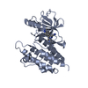
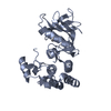
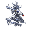
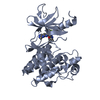
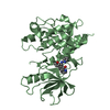
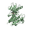
[English] 日本語
Yorodumi
- PDB-3psb: Furo[2,3-c]pyridine-based Indanone Oximes as Potent and Selective... -

+
Open data


ID or keywords:

Loading...

-
Basic information

Entry
Database: PDB / ID: 3psb
TitleFuro[2,3-c]pyridine-based Indanone Oximes as Potent and Selective B-Raf Inhibitors
ComponentsB-RAF PROTO-ONCOGENE SERINE/THREONINE-PROTEIN KINASE
KeywordsTRANSFERASE/TRANSFERASE INHIBITOR / Kinase / ATP-binding / Cardiomyopathy / Disease mutation / Metal-binding / Nucleotide-binding / Phorbol-ester binding / Phosphoprotein / Proto-oncogene / Serine/threonine-protein kinase / Zinc-finger / TRANSFERASE-TRANSFERASE INHIBITOR complex
Function / homology
Function and homology information


trehalose metabolism in response to stress / CD4-positive, alpha-beta T cell differentiation / CD4-positive or CD8-positive, alpha-beta T cell lineage commitment / negative regulation of synaptic vesicle exocytosis / head morphogenesis / Signalling to p38 via RIT and RIN / myeloid progenitor cell differentiation / ARMS-mediated activation / SHOC2 M1731 mutant abolishes MRAS complex function / Gain-of-function MRAS complexes activate RAF signaling ...trehalose metabolism in response to stress / CD4-positive, alpha-beta T cell differentiation / CD4-positive or CD8-positive, alpha-beta T cell lineage commitment / negative regulation of synaptic vesicle exocytosis / head morphogenesis / Signalling to p38 via RIT and RIN / myeloid progenitor cell differentiation / ARMS-mediated activation / SHOC2 M1731 mutant abolishes MRAS complex function / Gain-of-function MRAS complexes activate RAF signaling / endothelial cell apoptotic process / negative regulation of fibroblast migration / positive regulation of glucose transmembrane transport / establishment of protein localization to membrane / mitogen-activated protein kinase kinase binding / regulation of T cell differentiation / Negative feedback regulation of MAPK pathway / positive regulation of axonogenesis / Frs2-mediated activation / stress fiber assembly / positive regulation of axon regeneration / face development / synaptic vesicle exocytosis / somatic stem cell population maintenance / thyroid gland development / MAP kinase kinase activity / MAP kinase kinase kinase activity / negative regulation of endothelial cell apoptotic process / positive regulation of substrate adhesion-dependent cell spreading / response to cAMP / positive regulation of stress fiber assembly / ERK1 and ERK2 cascade / cellular response to calcium ion / substrate adhesion-dependent cell spreading / cellular response to nerve growth factor stimulus / thymus development / long-term synaptic potentiation / animal organ morphogenesis / Spry regulation of FGF signaling / RAF activation / Signaling by high-kinase activity BRAF mutants / visual learning / MAP2K and MAPK activation / epidermal growth factor receptor signaling pathway / response to peptide hormone / Negative regulation of MAPK pathway / Signaling by RAF1 mutants / Signaling by moderate kinase activity BRAF mutants / Paradoxical activation of RAF signaling by kinase inactive BRAF / Signaling downstream of RAS mutants / MAPK cascade / Signaling by BRAF and RAF1 fusions / cellular response to xenobiotic stimulus / presynapse / positive regulation of peptidyl-serine phosphorylation / T cell differentiation in thymus / cell body / regulation of cell population proliferation / T cell receptor signaling pathway / scaffold protein binding / negative regulation of neuron apoptotic process / positive regulation of ERK1 and ERK2 cascade / non-specific serine/threonine protein kinase / protein kinase activity / neuron projection / protein phosphorylation / protein serine kinase activity / intracellular membrane-bounded organelle / protein serine/threonine kinase activity / calcium ion binding / protein-containing complex binding / positive regulation of gene expression / negative regulation of apoptotic process / mitochondrion / ATP binding / identical protein binding / nucleus / plasma membrane / cytosol
Similarity search - Function
Raf-like Ras-binding domain / Raf-like Ras-binding / Ras-binding domain (RBD) profile. / Raf-like Ras-binding domain / Diacylglycerol/phorbol-ester binding / Phorbol esters/diacylglycerol binding domain (C1 domain) / Zinc finger phorbol-ester/DAG-type signature. / Zinc finger phorbol-ester/DAG-type profile. / Protein kinase C conserved region 1 (C1) domains (Cysteine-rich domains) / Protein kinase C-like, phorbol ester/diacylglycerol-binding domain ...Raf-like Ras-binding domain / Raf-like Ras-binding / Ras-binding domain (RBD) profile. / Raf-like Ras-binding domain / Diacylglycerol/phorbol-ester binding / Phorbol esters/diacylglycerol binding domain (C1 domain) / Zinc finger phorbol-ester/DAG-type signature. / Zinc finger phorbol-ester/DAG-type profile. / Protein kinase C conserved region 1 (C1) domains (Cysteine-rich domains) / Protein kinase C-like, phorbol ester/diacylglycerol-binding domain / C1-like domain superfamily / Protein tyrosine and serine/threonine kinase / Serine-threonine/tyrosine-protein kinase, catalytic domain / Ubiquitin-like domain superfamily / Transferase(Phosphotransferase) domain 1 / Transferase(Phosphotransferase); domain 1 / Phosphorylase Kinase; domain 1 / Phosphorylase Kinase; domain 1 / Serine/threonine-protein kinase, active site / Serine/Threonine protein kinases active-site signature. / Serine/Threonine protein kinases, catalytic domain / Protein kinase, ATP binding site / Protein kinases ATP-binding region signature. / Protein kinase domain profile. / Protein kinase domain / Protein kinase-like domain superfamily / 2-Layer Sandwich / Orthogonal Bundle / Mainly Alpha / Alpha Beta
Similarity search - Domain/homology
Chem-SM6 / Serine/threonine-protein kinase B-raf
Similarity search - Component
Biological speciesHomo sapiens (human)
MethodX-RAY DIFFRACTION / MOLECULAR REPLACEMENT / Resolution: 3.4 Å
AuthorsMorales, T. / Vigers, G.P.A. / Brandhuber, B.J.
CitationJournal: Bioorg.Med.Chem.Lett. / Year: 2011
Title: The Discovery of furo[2,3-c]pyridine-based indanone oximes as potent and selective B-Raf inhibitors.
Authors: Buckmelter, A.J. / Ren, L. / Laird, E.R. / Rast, B. / Miknis, G. / Wenglowsky, S. / Schlachter, S. / Welch, M. / Tarlton, E. / Grina, J. / Lyssikatos, J. / Brandhuber, B.J. / Morales, T. / ...Authors: Buckmelter, A.J. / Ren, L. / Laird, E.R. / Rast, B. / Miknis, G. / Wenglowsky, S. / Schlachter, S. / Welch, M. / Tarlton, E. / Grina, J. / Lyssikatos, J. / Brandhuber, B.J. / Morales, T. / Randolph, N. / Vigers, G. / Martinson, M. / Callejo, M.
History
DepositionDec 1, 2010Deposition site: RCSB / Processing site: RCSB
Revision 1.0Jan 19, 2011Provider: repository / Type: Initial release
Revision 1.1Jul 13, 2011Group: Version format compliance
Revision 1.2Jan 24, 2018Group: Structure summary / Category: audit_author / Item: _audit_author.name
Revision 1.3Sep 6, 2023Group: Data collection / Database references ...Data collection / Database references / Derived calculations / Refinement description
Category: chem_comp_atom / chem_comp_bond ...chem_comp_atom / chem_comp_bond / database_2 / pdbx_initial_refinement_model / struct_ref_seq_dif / struct_site
Item: _database_2.pdbx_DOI / _database_2.pdbx_database_accession ..._database_2.pdbx_DOI / _database_2.pdbx_database_accession / _struct_ref_seq_dif.details / _struct_site.pdbx_auth_asym_id / _struct_site.pdbx_auth_comp_id / _struct_site.pdbx_auth_seq_id

-
Structure visualization

Structure viewerMolecule:
MolmilJmol/JSmol

Downloads & links

-
Assembly

Deposited unit
A: B-RAF PROTO-ONCOGENE SERINE/THREONINE-PROTEIN KINASE
B: B-RAF PROTO-ONCOGENE SERINE/THREONINE-PROTEIN KINASE
hetero molecules


Theoretical massNumber of molelcules
Total (without water)71,1704
Polymers70,4352
Non-polymers7352
Water0
1
A: B-RAF PROTO-ONCOGENE SERINE/THREONINE-PROTEIN KINASE
hetero molecules


Theoretical massNumber of molelcules
Total (without water)35,5852
Polymers35,2181
Non-polymers3671
Water0
TypeNameSymmetry operationNumber
identity operation1_555x,y,z1
MethodPISA
2
B: B-RAF PROTO-ONCOGENE SERINE/THREONINE-PROTEIN KINASE
hetero molecules


Theoretical massNumber of molelcules
Total (without water)35,5852
Polymers35,2181
Non-polymers3671
Water0
TypeNameSymmetry operationNumber
identity operation1_555x,y,z1
MethodPISA
Unit cell
Length a, b, c (Å)101.073, 101.073, 162.952
Angle α, β, γ (deg.)90.00, 90.00, 90.00
Int Tables number92
Space group name H-MP41212

-
Components

#1: Protein B-RAF PROTO-ONCOGENE SERINE/THREONINE-PROTEIN KINASE / p94 / v-Raf murine sarcoma viral oncogene homolog B1


Mass: 35217.633 Da / Num. of mol.: 2 / Fragment: UNP residues 432-726
Source method: isolated from a genetically manipulated source
Source: (gene. exp.) Homo sapiens (human) / Gene: BRAF, BRAF1, RAFB1 / Production host: Spodoptera frugiperda (fall armyworm)
References: UniProt: P15056, non-specific serine/threonine protein kinase
#2: Chemical ChemComp-SM6 / ethyl 3-{[1-(hydroxyamino)-2H-inden-5-yl]amino}thieno[2,3-c]pyridine-2-carboxylate


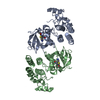
Mass: 367.422 Da / Num. of mol.: 2 / Source method: obtained synthetically / Formula: C19H17N3O3S

-
Experimental details

-
Experiment

ExperimentMethod: X-RAY DIFFRACTION / Number of used crystals: 1

-
Sample preparation

CrystalDensity Matthews: 2.95 Å3/Da / Density % sol: 58.36 %
Crystal growTemperature: 293 K / Method: vapor diffusion, hanging drop / pH: 8
Details: 10% PEG 8K 100mM Hepes pH8.0 3% aminocaproic acid 10% glycerol 2.6mg/ml protein 500uM inhibitor, VAPOR DIFFUSION, HANGING DROP, temperature 293K

-
Data collection

DiffractionMean temperature: 100 K
Diffraction sourceSource: ROTATING ANODE / Type: RIGAKU FR-E SUPERBRIGHT / Wavelength: 1.5418 Å
DetectorType: RIGAKU RAXIS IV++ / Detector: IMAGE PLATE / Date: Apr 29, 2005
RadiationMonochromator: Mirrors / Protocol: SINGLE WAVELENGTH / Monochromatic (M) / Laue (L): M / Scattering type: x-ray
Radiation wavelengthWavelength: 1.5418 Å / Relative weight: 1
ReflectionResolution: 3.4→30 Å / Num. all: 12196 / Num. obs: 12160 / % possible obs: 99.7 % / Observed criterion σ(I): 0 / Redundancy: 14.1 % / Rmerge(I) obs: 0.17 / Rsym value: 0.17 / Net I/σ(I): 6.12
Reflection shellResolution: 3.4→3.6 Å / Redundancy: 14.5 % / Rmerge(I) obs: 0.395 / Mean I/σ(I) obs: 2 / Num. unique all: 1727 / Rsym value: 0.381 / % possible all: 99.7

-
Processing

Software
NameVersionClassification
CrystalCleardata collection
MOLREPphasing
REFMAC5.5.0109refinement
MOSFLMdata reduction
SCALAdata scaling
RefinementMethod to determine structure: MOLECULAR REPLACEMENT
Starting model: 3D4Q
Resolution: 3.4→28.63 Å / Cor.coef. Fo:Fc: 0.897 / Cor.coef. Fo:Fc free: 0.871 / SU B: 25.508 / SU ML: 0.411 / Cross valid method: THROUGHOUT / ESU R Free: 0.564 / Stereochemistry target values: MAXIMUM LIKELIHOOD
RfactorNum. reflection% reflectionSelection details
Rfree0.2681 621 5.1 %RANDOM
Rwork0.22445 ---
obs0.22671 11511 100 %-
all-12196 --
Solvent computationIon probe radii: 0.8 Å / Shrinkage radii: 0.8 Å / VDW probe radii: 1.4 Å / Solvent model: BABINET MODEL WITH MASK
Displacement parametersBiso mean: 45.714 Å2
Baniso -1Baniso -2Baniso -3
1-0.04 Å20 Å20 Å2
2--0.04 Å20 Å2
3----0.09 Å2
Refinement stepCycle: LAST / Resolution: 3.4→28.63 Å
ProteinNucleic acidLigandSolventTotal
Num. atoms4200 0 52 0 4252
Refine LS restraints
Refine-IDTypeDev idealDev ideal targetNumber
X-RAY DIFFRACTIONr_bond_refined_d0.0070.0224350
X-RAY DIFFRACTIONr_bond_other_d
X-RAY DIFFRACTIONr_angle_refined_deg1.1361.9785877
X-RAY DIFFRACTIONr_angle_other_deg
X-RAY DIFFRACTIONr_dihedral_angle_1_deg5.4685522
X-RAY DIFFRACTIONr_dihedral_angle_2_deg36.53223.871186
X-RAY DIFFRACTIONr_dihedral_angle_3_deg18.12515793
X-RAY DIFFRACTIONr_dihedral_angle_4_deg16.1981526
X-RAY DIFFRACTIONr_chiral_restr0.0750.2645
X-RAY DIFFRACTIONr_gen_planes_refined0.0040.0213248
X-RAY DIFFRACTIONr_gen_planes_other
X-RAY DIFFRACTIONr_nbd_refined
X-RAY DIFFRACTIONr_nbd_other
X-RAY DIFFRACTIONr_nbtor_refined
X-RAY DIFFRACTIONr_nbtor_other
X-RAY DIFFRACTIONr_xyhbond_nbd_refined
X-RAY DIFFRACTIONr_xyhbond_nbd_other
X-RAY DIFFRACTIONr_metal_ion_refined
X-RAY DIFFRACTIONr_metal_ion_other
X-RAY DIFFRACTIONr_symmetry_vdw_refined
X-RAY DIFFRACTIONr_symmetry_vdw_other
X-RAY DIFFRACTIONr_symmetry_hbond_refined
X-RAY DIFFRACTIONr_symmetry_hbond_other
X-RAY DIFFRACTIONr_symmetry_metal_ion_refined
X-RAY DIFFRACTIONr_symmetry_metal_ion_other
X-RAY DIFFRACTIONr_mcbond_it0.3891.52616
X-RAY DIFFRACTIONr_mcbond_other
X-RAY DIFFRACTIONr_mcangle_it0.71324236
X-RAY DIFFRACTIONr_scbond_it0.51831734
X-RAY DIFFRACTIONr_scangle_it0.9434.51641
X-RAY DIFFRACTIONr_rigid_bond_restr
X-RAY DIFFRACTIONr_sphericity_free
X-RAY DIFFRACTIONr_sphericity_bonded
LS refinement shellResolution: 3.4→3.487 Å / Total num. of bins used: 20
RfactorNum. reflection% reflection
Rfree0.32 43 -
Rwork0.264 832 -
obs--100 %

+
About Yorodumi

-
News

-
Feb 9, 2022. New format data for meta-information of EMDB entries

New format data for meta-information of EMDB entries

  • Version 3 of the EMDB header file is now the official format.
  • The previous official version 1.9 will be removed from the archive.

Related info.:EMDB header

External links:wwPDB to switch to version 3 of the EMDB data model

-
Aug 12, 2020. Covid-19 info

Covid-19 info

URL: https://pdbj.org/emnavi/covid19.php

New page: Covid-19 featured information page in EM Navigator.

Related info.:Covid-19 info / Mar 5, 2020. Novel coronavirus structure data

+
Mar 5, 2020. Novel coronavirus structure data

Novel coronavirus structure data

Related info.:Yorodumi Speices / Aug 12, 2020. Covid-19 info

External links:COVID-19 featured content - PDBj / Molecule of the Month (242):Coronavirus Proteases

+
Jan 31, 2019. EMDB accession codes are about to change! (news from PDBe EMDB page)

EMDB accession codes are about to change! (news from PDBe EMDB page)

  • The allocation of 4 digits for EMDB accession codes will soon come to an end. Whilst these codes will remain in use, new EMDB accession codes will include an additional digit and will expand incrementally as the available range of codes is exhausted. The current 4-digit format prefixed with “EMD-” (i.e. EMD-XXXX) will advance to a 5-digit format (i.e. EMD-XXXXX), and so on. It is currently estimated that the 4-digit codes will be depleted around Spring 2019, at which point the 5-digit format will come into force.
  • The EM Navigator/Yorodumi systems omit the EMD- prefix.

Related info.:Q: What is EMD? / ID/Accession-code notation in Yorodumi/EM Navigator

External links:EMDB Accession Codes are Changing Soon! / Contact to PDBj

+
Jul 12, 2017. Major update of PDB

Major update of PDB

  • wwPDB released updated PDB data conforming to the new PDBx/mmCIF dictionary.
  • This is a major update changing the version number from 4 to 5, and with Remediation, in which all the entries are updated.
  • In this update, many items about electron microscopy experimental information are reorganized (e.g. em_software).
  • Now, EM Navigator and Yorodumi are based on the updated data.

External links:wwPDB Remediation / Enriched Model Files Conforming to OneDep Data Standards Now Available in the PDB FTP Archive

-
Yorodumi

Thousand views of thousand structures

  • Yorodumi is a browser for structure data from EMDB, PDB, SASBDB, etc.
  • This page is also the successor to EM Navigator detail page, and also detail information page/front-end page for Omokage search.
  • The word "yorodu" (or yorozu) is an old Japanese word meaning "ten thousand". "mi" (miru) is to see.

Related info.:EMDB / PDB / SASBDB / Comparison of 3 databanks / Yorodumi Search / Aug 31, 2016. New EM Navigator & Yorodumi / Yorodumi Papers / Jmol/JSmol / Function and homology information / Changes in new EM Navigator and Yorodumi

Read more