 Movie
Movie Controller


+ Open data

- Basic information
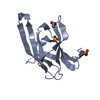
| Entry | Database: PDB / ID: 1gd3 | ||||||
|---|---|---|---|---|---|---|---|
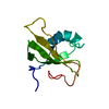
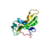
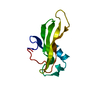
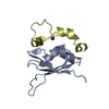
| Title | refined solution structure of human cystatin A | ||||||
 Components Components | CYSTATIN A | ||||||
 Keywords Keywords | PROTEIN BINDING / cystatin A / Thiol protease inhibitor | ||||||
| Function / homology |  Function and homology information Function and homology informationnegative regulation of peptidase activity / peptidase inhibitor complex / Formation of the cornified envelope / peptide cross-linking / cornified envelope / cysteine-type endopeptidase inhibitor activity / keratinocyte differentiation / negative regulation of proteolysis / cell-cell adhesion / protease binding ...negative regulation of peptidase activity / peptidase inhibitor complex / Formation of the cornified envelope / peptide cross-linking / cornified envelope / cysteine-type endopeptidase inhibitor activity / keratinocyte differentiation / negative regulation of proteolysis / cell-cell adhesion / protease binding / extracellular space / nucleoplasm / cytosol / cytoplasmSimilarity search - Function | ||||||
| Biological species |  Homo sapiens (human) Homo sapiens (human) | ||||||
| Method | SOLUTION NMR / distance geometry, simulated annealing | ||||||
 Authors Authors | Shimba, N. / Kariya, E. / Tate, S. / Kaji, H. / Kainosho, M. | ||||||
 Citation Citation | Journal: J.STRUCT.FUNCT.GENOM. / Year: 2000 Title: Structural comparison between wild-type and P25S human cystatin A by NMR spectroscopy. Does this mutation affect the a-helix conformation ? Authors: Shimba, N. / Kariya, E. / Tate, S. / Kaji, H. / Kainosho, M. | ||||||
| History |
|
- Structure visualization
| Structure viewer | Molecule: MolmilJmol/JSmol |
|---|
- Downloads & links
-Download
| PDBx/mmCIF format | 1gd3.cif.gz | 43.8 KB | Display | PDBx/mmCIF format |
|---|---|---|---|---|
| PDB format | pdb1gd3.ent.gz | 32.1 KB | Display | PDB format |
| PDBx/mmJSON format | 1gd3.json.gz | Tree view | PDBx/mmJSON format | |
| Others |  Other downloads Other downloads |
-Validation report
| Arichive directory | https://data.pdbj.org/pub/pdb/validation_reports/gd/1gd3ftp://data.pdbj.org/pub/pdb/validation_reports/gd/1gd3 | HTTPS FTP |
|---|
-Related structure data


-Links
 PDBj
PDBj
- Assembly
| Deposited unit | 
| |||||||||
|---|---|---|---|---|---|---|---|---|---|---|
| 1 |
| |||||||||
| NMR ensembles |
|
-Components
| #1: Protein | Mass: 11002.426 Da / Num. of mol.: 1 / Mutation: M65L Source method: isolated from a genetically manipulated source Source: (gene. exp.) Homo sapiens (human) / Production host:  Escherichia coli (E. coli) / References: UniProt: P01040 Escherichia coli (E. coli) / References: UniProt: P01040 |
|---|
-Experimental details
-Experiment
| Experiment | Method: SOLUTION NMR | ||||||||||||||||
|---|---|---|---|---|---|---|---|---|---|---|---|---|---|---|---|---|---|
| NMR experiment |
| ||||||||||||||||
| NMR details | Text: The structure was determined using triple-resonance NMR spectroscopy. |
- Sample preparation
| Details |
| ||||||||||||
|---|---|---|---|---|---|---|---|---|---|---|---|---|---|
| Sample conditions | Ionic strength: 0 / pH: 4.0 / Pressure: ambient / Temperature: 310 K | ||||||||||||
| Crystal grow | *PLUS Method: other / Details: NMR |
-NMR measurement
| NMR spectrometer | Type: Bruker DRX / Manufacturer: Bruker / Model: DRX / Field strength: 800 MHz |
|---|
- Processing
| NMR software | Name: X-PLOR / Version: 3.85 / Classification: refinement |
|---|---|
| Refinement | Method: distance geometry, simulated annealing / Software ordinal: 1 |
| NMR ensemble | Conformers submitted total number: 1 |
