 Movie
Movie Controller

[English] 日本語
 Yorodumi
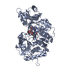
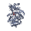
- PDB-1g0i: CRYSTAL STRUCTURE OF MJ0109 GENE PRODUCT INOSITOL MONOPHOSPHATASE... -
+ Open data

- Basic information
| Entry | Database: PDB / ID: 1g0i | ||||||
|---|---|---|---|---|---|---|---|
| Title | CRYSTAL STRUCTURE OF MJ0109 GENE PRODUCT INOSITOL MONOPHOSPHATASE-FRUCTOSE 1,6 BISPHOSPHATASE | ||||||
 Components Components | INOSITOL MONOPHOSPHATASE Inositol-phosphate phosphatase Inositol-phosphate phosphatase | ||||||
 Keywords Keywords | HYDROLASE / HOMODIMER / COMPLEXED WITH Mn2+ / inositol / and phosphate | ||||||
| Function / homology |  Function and homology informationinositol-phosphate phosphatase / inositol monophosphate 3-phosphatase activity / inositol monophosphate 4-phosphatase activity / inositol monophosphate 1-phosphatase activity / inositol metabolic process / fructose-bisphosphatase / fructose 1,6-bisphosphate 1-phosphatase activity / phosphatidylinositol phosphate biosynthetic process / carbohydrate metabolic process / signal transduction / metal ion binding Function and homology informationinositol-phosphate phosphatase / inositol monophosphate 3-phosphatase activity / inositol monophosphate 4-phosphatase activity / inositol monophosphate 1-phosphatase activity / inositol metabolic process / fructose-bisphosphatase / fructose 1,6-bisphosphate 1-phosphatase activity / phosphatidylinositol phosphate biosynthetic process / carbohydrate metabolic process / signal transduction / metal ion bindingSimilarity search - Function | ||||||
| Biological species |   Methanocaldococcus jannaschii (archaea) Methanocaldococcus jannaschii (archaea) | ||||||
| Method | X-RAY DIFFRACTION / Resolution: 2.4 Å | ||||||
 Authors Authors | Johnson, K.A. / Chen, L. / Yang, H. / Roberts, M.F. / Stec, B. | ||||||
 Citation Citation | Journal: Biochemistry / Year: 2001 Title: Crystal structure and catalytic mechanism of the MJ0109 gene product: a bifunctional enzyme with inositol monophosphatase and fructose 1,6-bisphosphatase activities. Authors: Johnson, K.A. / Chen, L. / Yang, H. / Roberts, M.F. / Stec, B. #1: Journal: Nat.Struct.Biol. / Year: 2000Title: MJ0109 is an Enzyme that is Both an Inositol Monophosphatse and the 'Missing' Archaeal Fructose-1,6-Bisphosphatase Authors: Stec, B. / Yang, H. / Johnson, K.A. / Chen, L. / Roberts, M.F. | ||||||
| History |
|
- Structure visualization
| Structure viewer | Molecule: MolmilJmol/JSmol |
|---|
- Downloads & links
-Download
| PDBx/mmCIF format | 1g0i.cif.gz | 115.7 KB | Display | PDBx/mmCIF format |
|---|---|---|---|---|
| PDB format | pdb1g0i.ent.gz | 89.2 KB | Display | PDB format |
| PDBx/mmJSON format | 1g0i.json.gz | Tree view | PDBx/mmJSON format | |
| Others |  Other downloads Other downloads |
-Validation report
| Arichive directory | https://data.pdbj.org/pub/pdb/validation_reports/g0/1g0iftp://data.pdbj.org/pub/pdb/validation_reports/g0/1g0i | HTTPS FTP |
|---|
-Related structure data
| Related structure data |  1g0hC 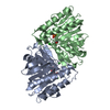 1awbS C: citing same article ( S: Starting model for refinement |
|---|---|
| Similar structure data |


-Links
 PDBj
PDBj- Assembly
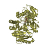
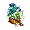
| Deposited unit | 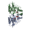
| ||||||||
|---|---|---|---|---|---|---|---|---|---|
| 1 |
| ||||||||
| Unit cell |
| ||||||||
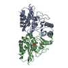
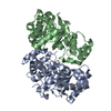
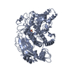
| Details | The biological assembly is a dimer constructed from chain A and B |
-Components
| #1: Protein | Inositol-phosphate phosphatase / MJ0109 GENE PRODUCT Mass: 28611.838 Da / Num. of mol.: 2 Source method: isolated from a genetically manipulated source Source: (gene. exp.) Methanocaldococcus jannaschii (archaea)Plasmid: PET23A+ / Production host:  Escherichia coli (E. coli) / References: UniProt: Q57573, inositol-phosphate phosphatase Escherichia coli (E. coli) / References: UniProt: Q57573, inositol-phosphate phosphatase#2: Chemical | ChemComp-MN /   Mass: 54.938 Da / Num. of mol.: 6 / Source method: obtained synthetically / Formula: Mn Mass: 54.938 Da / Num. of mol.: 6 / Source method: obtained synthetically / Formula: Mn#3: Chemical | Phosphate  Mass: 94.971 Da / Num. of mol.: 2 / Source method: obtained synthetically / Formula: PO4 Mass: 94.971 Da / Num. of mol.: 2 / Source method: obtained synthetically / Formula: PO4#4: Chemical | Inositol  Mass: 180.156 Da / Num. of mol.: 2 / Source method: obtained synthetically / Formula: C6H12O6 / Comment: neurotransmitter, hormone*YM Mass: 180.156 Da / Num. of mol.: 2 / Source method: obtained synthetically / Formula: C6H12O6 / Comment: neurotransmitter, hormone*YM#5: Water | ChemComp-HOH / | Water Mass: 18.015 Da / Num. of mol.: 48 / Source method: isolated from a natural source / Formula: H2O Mass: 18.015 Da / Num. of mol.: 48 / Source method: isolated from a natural source / Formula: H2O |
|---|
-Experimental details
-Experiment
| Experiment | Method: X-RAY DIFFRACTION / Number of used crystals: 1 |
|---|
- Sample preparation
| Crystal | Density Matthews: 3.03 Å3/Da / Density % sol: 59.42 % | ||||||||||||||||||||||||||||||||||||||||||
|---|---|---|---|---|---|---|---|---|---|---|---|---|---|---|---|---|---|---|---|---|---|---|---|---|---|---|---|---|---|---|---|---|---|---|---|---|---|---|---|---|---|---|---|
| Crystal grow | Temperature: 298 K / Method: vapor diffusion, hanging drop / pH: 7.2 Details: PEG 8000, sodium chloride, manganese chloride, TRIS, pH 7.2, VAPOR DIFFUSION, HANGING DROP, temperature 298.0K | ||||||||||||||||||||||||||||||||||||||||||
| Crystal grow | *PLUS | ||||||||||||||||||||||||||||||||||||||||||
| Components of the solutions | *PLUS
|
-Data collection
| Diffraction | Mean temperature: 298 K |
|---|---|
| Diffraction source | Source: ROTATING ANODE / Type: RIGAKU RU200 / Wavelength: 1.5418 |
| Detector | Type: SDMS / Detector: AREA DETECTOR / Date: Jul 26, 1997 |
| Radiation | Monochromator: GRAPHITE / Protocol: SINGLE WAVELENGTH / Monochromatic (M) / Laue (L): M / Scattering type: x-ray |
| Radiation wavelength | Wavelength: 1.5418 Å / Relative weight: 1 |
| Reflection | Resolution: 2.4→30 Å / Num. all: 26181 / Num. obs: 25134 / % possible obs: 92.4 % / Observed criterion σ(F): 0 / Observed criterion σ(I): 0 / Redundancy: 3 % / Biso Wilson estimate: 38.8 Å2 / Rmerge(I) obs: 0.09 / Net I/σ(I): 7.5 |
| Reflection shell | Resolution: 2.4→2.5 Å / Redundancy: 2.1 % / Rmerge(I) obs: 0.41 / Mean I/σ(I) obs: 1 / Num. unique all: 2758 / % possible all: 90.5 |
| Reflection | *PLUS Num. obs: 26224 / % possible obs: 97.4 % / Rmerge(I) obs: 0.09 |
| Reflection shell | *PLUS % possible obs: 94.5 % |
- Processing
| Software |
| |||||||||||||||||||||||||
|---|---|---|---|---|---|---|---|---|---|---|---|---|---|---|---|---|---|---|---|---|---|---|---|---|---|---|
| Refinement | Starting model: 1AWB Resolution: 2.4→12 Å / Num. parameters: 16495 / Num. restraintsaints: 16934 / σ(F): 0 / σ(I): 0 / Stereochemistry target values: Engh & Huber / Details: conjugated gradient least squares
| |||||||||||||||||||||||||
| Solvent computation | Solvent model: MOEWS & KRETSINGER | |||||||||||||||||||||||||
| Refine analyze | Num. disordered residues: 0 | |||||||||||||||||||||||||
| Refinement step | Cycle: LAST / Resolution: 2.4→12 Å
| |||||||||||||||||||||||||
| Refine LS restraints |
| |||||||||||||||||||||||||
| Software | *PLUS Name: SHELXL-97 / Classification: refinement | |||||||||||||||||||||||||
| Refinement | *PLUS Highest resolution: 2.4 Å / Lowest resolution: 12 Å / σ(F): 0 / Rfactor all: 0.251 / Rfactor obs: 0.192 / Rfactor Rfree: 0.322 | |||||||||||||||||||||||||
| Solvent computation | *PLUS | |||||||||||||||||||||||||
| Displacement parameters | *PLUS | |||||||||||||||||||||||||
| Refine LS restraints | *PLUS Type: s_angle_d / Dev ideal: 0.021 |
