 Movie
Movie Controller

[English] 日本語
 Yorodumi
- PDB-1fxh: MUTANT OF PENICILLIN ACYLASE IMPAIRED IN CATALYSIS WITH PHENYLACE... -
+ Open data

- Basic information
| Entry | Database: PDB / ID: 1fxh | ||||||
|---|---|---|---|---|---|---|---|
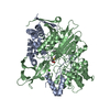
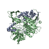
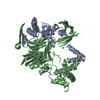
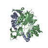
| Title | MUTANT OF PENICILLIN ACYLASE IMPAIRED IN CATALYSIS WITH PHENYLACETIC ACID IN THE ACTIVE SITE | ||||||
 Components Components | (PENICILLIN ACYLASE Penicillin amidase) x 2 Penicillin amidase) x 2 | ||||||
 Keywords Keywords | HYDROLASE / Ntn-hydrolase fold | ||||||
| Function / homology |  Function and homology informationpenicillin amidase / penicillin amidase activity / antibiotic biosynthetic process / periplasmic space / response to antibiotic / metal ion binding Function and homology informationpenicillin amidase / penicillin amidase activity / antibiotic biosynthetic process / periplasmic space / response to antibiotic / metal ion bindingSimilarity search - Function | ||||||
| Biological species |  Escherichia coli (E. coli) Escherichia coli (E. coli) | ||||||
| Method | X-RAY DIFFRACTION / SYNCHROTRON / Resolution: 1.97 Å | ||||||
 Authors Authors | Alkema, W.B. / Hensgens, C.M. / Kroezinga, E.H. / de Vries, E. / Floris, R. / van der Laan, J.M. / Dijkstra, B.W. / Janssen, D.B. | ||||||
 Citation Citation | Journal: Protein Eng. / Year: 2000 Title: Characterization of the beta-lactam binding site of penicillin acylase of Escherichia coli by structural and site-directed mutagenesis studies. Authors: Alkema, W.B. / Hensgens, C.M. / Kroezinga, E.H. / de Vries, E. / Floris, R. / van der Laan, J.M. / Dijkstra, B.W. / Janssen, D.B. | ||||||
| History |
|
- Structure visualization
| Structure viewer | Molecule: MolmilJmol/JSmol |
|---|
- Downloads & links
-Download
| PDBx/mmCIF format | 1fxh.cif.gz | 178.7 KB | Display | PDBx/mmCIF format |
|---|---|---|---|---|
| PDB format | pdb1fxh.ent.gz | 137 KB | Display | PDB format |
| PDBx/mmJSON format | 1fxh.json.gz | Tree view | PDBx/mmJSON format | |
| Others |  Other downloads Other downloads |
-Validation report
| Arichive directory | https://data.pdbj.org/pub/pdb/validation_reports/fx/1fxhftp://data.pdbj.org/pub/pdb/validation_reports/fx/1fxh | HTTPS FTP |
|---|
-Related structure data


-Links
 PDBj
PDBj


- Assembly
| Deposited unit | 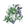
| ||||||||
|---|---|---|---|---|---|---|---|---|---|
| 1 |
| ||||||||
| Unit cell |
| ||||||||
| Details | The biological assembly is a hetero dimer. |
-Components
| #1: Protein | Penicillin amidase Mass: 23838.824 Da / Num. of mol.: 1 / Fragment: ALPHA SUBUNIT Source method: isolated from a genetically manipulated source Source: (gene. exp.) Escherichia coli (E. coli) / Plasmid: PEC / Production host: Escherichia coli (E. coli) / References: UniProt: P06875, penicillin amidase |
|---|---|
| #2: Protein | Penicillin amidase Mass: 62400.496 Da / Num. of mol.: 1 / Fragment: BETA SUBUNIT / Mutation: N241A, V148L Source method: isolated from a genetically manipulated source Source: (gene. exp.) Escherichia coli (E. coli) / Plasmid: PEC / Production host: Escherichia coli (E. coli) / References: UniProt: P06875, penicillin amidase |
| #3: Chemical | ChemComp-CA /   Mass: 40.078 Da / Num. of mol.: 1 / Source method: obtained synthetically / Formula: Ca Mass: 40.078 Da / Num. of mol.: 1 / Source method: obtained synthetically / Formula: Ca |
| #4: Chemical | ChemComp-PAC / Phenylacetic acid  Mass: 136.148 Da / Num. of mol.: 1 / Source method: obtained synthetically / Formula: C8H8O2 Mass: 136.148 Da / Num. of mol.: 1 / Source method: obtained synthetically / Formula: C8H8O2 |
| #5: Water | ChemComp-HOH / Water Mass: 18.015 Da / Num. of mol.: 653 / Source method: isolated from a natural source / Formula: H2O Mass: 18.015 Da / Num. of mol.: 653 / Source method: isolated from a natural source / Formula: H2O |
-Experimental details
-Experiment
| Experiment | Method: X-RAY DIFFRACTION / Number of used crystals: 1 |
|---|
- Sample preparation
| Crystal | Density Matthews: 2.18 Å3/Da / Density % sol: 43.59 % | ||||||||||||||||||||||||
|---|---|---|---|---|---|---|---|---|---|---|---|---|---|---|---|---|---|---|---|---|---|---|---|---|---|
| Crystal grow | Temperature: 277 K / Method: vapor diffusion, hanging drop / pH: 7.2 Details: PEG MME 2000, MOPS, pH 7.2, VAPOR DIFFUSION, HANGING DROP, temperature 277K | ||||||||||||||||||||||||
| Crystal grow | *PLUS Temperature: 4 ℃Details: drop consists of equal amounts of protein and precipitant solutions | ||||||||||||||||||||||||
| Components of the solutions | *PLUS
|
-Data collection
| Diffraction | Mean temperature: 100 K |
|---|---|
| Diffraction source | Source: SYNCHROTRON / Site: EMBL/DESY, HAMBURG  / Beamline: X31 / Wavelength: 1.04 / Beamline: X31 / Wavelength: 1.04 |
| Detector | Type: MARRESEARCH / Detector: IMAGE PLATE / Date: Sep 25, 1998 |
| Radiation | Protocol: SINGLE WAVELENGTH / Monochromatic (M) / Laue (L): M / Scattering type: x-ray |
| Radiation wavelength | Wavelength: 1.04 Å / Relative weight: 1 |
| Reflection | Resolution: 1.97→20 Å / Num. all: 51862 / Num. obs: 50404 / % possible obs: 97.3 % / Observed criterion σ(F): 0 / Observed criterion σ(I): 0 / Redundancy: 1.8 % / Biso Wilson estimate: 5.7 Å2 / Rmerge(I) obs: 0.041 / Net I/σ(I): 15.7 |
| Reflection shell | Resolution: 1.97→2 Å / Rmerge(I) obs: 0.124 / Num. unique all: 0 / % possible all: 95 |
| Reflection shell | *PLUS % possible obs: 95 % |
- Processing
| Software |
| |||||||||||||||||||||||||
|---|---|---|---|---|---|---|---|---|---|---|---|---|---|---|---|---|---|---|---|---|---|---|---|---|---|---|
| Refinement | Resolution: 1.97→20 Å / σ(F): 0 / σ(I): 0 / Stereochemistry target values: ENGH & HUBER / Details: USED MAXIMUM LIKELYHOOD
| |||||||||||||||||||||||||
| Refinement step | Cycle: LAST / Resolution: 1.97→20 Å
| |||||||||||||||||||||||||
| Refine LS restraints |
| |||||||||||||||||||||||||
| Software | *PLUS Name: CNS / Classification: refinement | |||||||||||||||||||||||||
| Refine LS restraints | *PLUS
|
