 Movie
Movie Controller

[English] 日本語
 Yorodumi
- PDB-1fqd: CRYSTAL STRUCTURE OF MALTOTETRAITOL BOUND TO CLOSED-FORM MALTODEX... -
+ Open data

- Basic information
| Entry | Database: PDB / ID: 1fqd | |||||||||
|---|---|---|---|---|---|---|---|---|---|---|
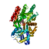
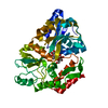
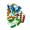
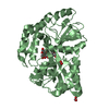
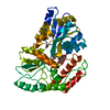
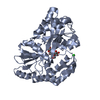
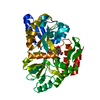
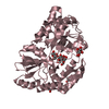
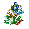
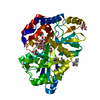
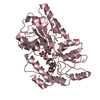
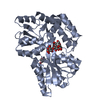
| Title | CRYSTAL STRUCTURE OF MALTOTETRAITOL BOUND TO CLOSED-FORM MALTODEXTRIN BINDING PROTEIN | |||||||||
 Components Components | MALTODEXTRIN-BINDING PROTEIN | |||||||||
 Keywords Keywords | SUGAR BINDING PROTEIN / sugar-binding protein / maltotetraitol | |||||||||
| Function / homology |  Function and homology information Function and homology informationdetection of maltose stimulus / maltose transport complex /  maltose binding / maltose transport / maltodextrin transmembrane transport / carbohydrate transport / carbohydrate transmembrane transporter activity / ATP-binding cassette (ABC) transporter complex, substrate-binding subunit-containing / ATP-binding cassette (ABC) transporter complex / cell chemotaxis ...detection of maltose stimulus / maltose transport complex / maltose binding / maltose transport / maltodextrin transmembrane transport / carbohydrate transport / carbohydrate transmembrane transporter activity / ATP-binding cassette (ABC) transporter complex, substrate-binding subunit-containing / ATP-binding cassette (ABC) transporter complex / cell chemotaxis / outer membrane-bounded periplasmic space / periplasmic space / DNA damage response / membrane maltose binding / maltose transport / maltodextrin transmembrane transport / carbohydrate transport / carbohydrate transmembrane transporter activity / ATP-binding cassette (ABC) transporter complex, substrate-binding subunit-containing / ATP-binding cassette (ABC) transporter complex / cell chemotaxis ...detection of maltose stimulus / maltose transport complex / maltose binding / maltose transport / maltodextrin transmembrane transport / carbohydrate transport / carbohydrate transmembrane transporter activity / ATP-binding cassette (ABC) transporter complex, substrate-binding subunit-containing / ATP-binding cassette (ABC) transporter complex / cell chemotaxis / outer membrane-bounded periplasmic space / periplasmic space / DNA damage response / membraneSimilarity search - Function | |||||||||
| Biological species |  Escherichia coli (E. coli) Escherichia coli (E. coli) | |||||||||
| Method | X-RAY DIFFRACTION / Resolution: 2.3 Å | |||||||||
 Authors Authors | Duan, X. / Hall, J.A. / Nikaido, H. / Quiocho, F.A. | |||||||||
 Citation Citation | Journal: J Mol Biol / Year: 2001 Title: Crystal structures of the maltodextrin/maltose-binding protein complexed with reduced oligosaccharides: flexibility of tertiary structure and ligand binding. Authors: X Duan / J A Hall / H Nikaido / F A Quiocho /  Abstract: The structure of the maltodextrin or maltose-binding protein, an initial receptor for bacterial ABC-type active transport and chemotaxis, consists of two globular domains that are separated by a ...The structure of the maltodextrin or maltose-binding protein, an initial receptor for bacterial ABC-type active transport and chemotaxis, consists of two globular domains that are separated by a groove wherein the ligand is bound and enclosed by an inter-domain rotation. Here, we report the determination of the crystal structures of the protein complexed with reduced maltooligosaccharides (maltotriitol and maltotetraitol) in both the "closed" and "open" forms. Although these modified sugars bind to the receptor, they are not transported by the wild-type transporter. In the closed structures, the reduced sugars are buried in the groove and bound by both domains, one domain mainly by hydrogen-bonding interactions and the other domain primarily by non-polar interactions with aromatic side-chains. In the open structures, which abrogate both cellular activities of active transport and chemotaxis because of the large separation between the two domains, the sugars are bound almost exclusively to the domain rich in aromatic residues. The binding site for the open chain glucitol residue extends to a subsite that is distinct from those for the glucose residues that were uncovered in prior structural studies of the binding of active linear maltooligosaccharides. Occupation of this subsite may also account for the inability of the reduced oligosaccharides to be transported. The structures reported here, combined with those previously determined for several other complexes with active oligosaccharides in the closed form and with cyclodextrin in the open form, revealed at least four distinct modes of ligand binding but with only one being functionally active. This versatility reflects the flexibility of the protein, from very large motions of interdomain rotation to more localized side-chain conformational changes, and adaptation by the oligosaccharides as well. | |||||||||
| History |
|
- Structure visualization
| Structure viewer | Molecule: MolmilJmol/JSmol |
|---|
- Downloads & links
-Download
| PDBx/mmCIF format | 1fqd.cif.gz | 86.9 KB | Display | PDBx/mmCIF format |
|---|---|---|---|---|
| PDB format | pdb1fqd.ent.gz | 65.5 KB | Display | PDB format |
| PDBx/mmJSON format | 1fqd.json.gz | Tree view | PDBx/mmJSON format | |
| Others |  Other downloads Other downloads |
-Validation report
| Arichive directory | https://data.pdbj.org/pub/pdb/validation_reports/fq/1fqdftp://data.pdbj.org/pub/pdb/validation_reports/fq/1fqd | HTTPS FTP |
|---|
-Related structure data


-Links
 PDBj
PDBj


- Assembly
| Deposited unit | 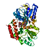
| ||||||||||
|---|---|---|---|---|---|---|---|---|---|---|---|
| 1 |
| ||||||||||
| Unit cell |
|
-Components
| #1: Protein | Mass: 40695.051 Da / Num. of mol.: 1 Source method: isolated from a genetically manipulated source Details: COMPLEXED WITH MALTOTETRAITOL / Source: (gene. exp.) Escherichia coli (E. coli) / Production host: Escherichia coli (E. coli) / References: UniProt: P02928, UniProt: P0AEX9*PLUS |
|---|---|
| #2: Polysaccharide | alpha-D-glucopyranose-(1-4)-alpha-D-glucopyranose-(1-4)-alpha-D-glucopyranose-(1-4)-D-glucose / Mass: 666.578 Da / Num. of mol.: 1 Source method: isolated from a genetically manipulated source |
| #3: Water | ChemComp-HOH / Water Mass: 18.015 Da / Num. of mol.: 105 / Source method: isolated from a natural source / Formula: H2O Mass: 18.015 Da / Num. of mol.: 105 / Source method: isolated from a natural source / Formula: H2O |
-Experimental details
-Experiment
| Experiment | Method: X-RAY DIFFRACTION / Number of used crystals: 1 |
|---|
- Sample preparation
| Crystal | Density Matthews: 2.31 Å3/Da / Density % sol: 46.83 % | ||||||||||||||||||||||||||||||||||||||||||||||||||
|---|---|---|---|---|---|---|---|---|---|---|---|---|---|---|---|---|---|---|---|---|---|---|---|---|---|---|---|---|---|---|---|---|---|---|---|---|---|---|---|---|---|---|---|---|---|---|---|---|---|---|---|
| Crystal grow | Temperature: 293 K / Method: vapor diffusion, hanging drop / pH: 6.2 Details: PEG 6000, Mes, NaN3, pH 6.2, VAPOR DIFFUSION, HANGING DROP, temperature 293K | ||||||||||||||||||||||||||||||||||||||||||||||||||
| Crystal grow | *PLUS | ||||||||||||||||||||||||||||||||||||||||||||||||||
| Components of the solutions | *PLUS
|
-Data collection
| Diffraction | Mean temperature: 200 K |
|---|---|
| Diffraction source | Source: ROTATING ANODE / Type: RIGAKU RU200 / Wavelength: 1.5418 |
| Detector | Type: MACSCIENCE / Detector: IMAGE PLATE |
| Radiation | Protocol: SINGLE WAVELENGTH / Monochromatic (M) / Laue (L): M / Scattering type: x-ray |
| Radiation wavelength | Wavelength: 1.5418 Å / Relative weight: 1 |
| Reflection | Resolution: 2.2→30 Å / % possible obs: 99.3 % / Observed criterion σ(F): 2 / Observed criterion σ(I): 1 / Rmerge(I) obs: 0.046 |
| Reflection shell | Resolution: 2.2→2.3 Å / Rmerge(I) obs: 0.143 / % possible all: 80 |
| Reflection | *PLUS Lowest resolution: 30 Å / Num. measured all: 92330 |
| Reflection shell | *PLUS % possible obs: 80 % |
- Processing
| Software |
| ||||||||||||||||
|---|---|---|---|---|---|---|---|---|---|---|---|---|---|---|---|---|---|
| Refinement | Resolution: 2.3→10 Å / Cross valid method: THROUGHOUT / σ(F): 2
| ||||||||||||||||
| Refinement step | Cycle: LAST / Resolution: 2.3→10 Å
| ||||||||||||||||
| Refine LS restraints |
| ||||||||||||||||
| Software | *PLUS Name: X-PLOR / Classification: refinement | ||||||||||||||||
| Refinement | *PLUS Highest resolution: 2.3 Å / Lowest resolution: 10 Å / σ(F): 2 / Rfactor obs: 0.191 | ||||||||||||||||
| Solvent computation | *PLUS | ||||||||||||||||
| Displacement parameters | *PLUS | ||||||||||||||||
| Refine LS restraints | *PLUS
|
