 Movie
Movie Controller


+ Open data

- Basic information
| Entry | Database: PDB / ID: 1f7w | ||||||
|---|---|---|---|---|---|---|---|
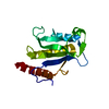
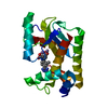
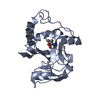
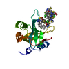
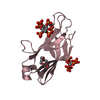
| Title | SOLUTION STRUCTURE OF C-TERMINAL DOMAIN ZIPA | ||||||
 Components Components | CELL DIVISION PROTEIN ZIPA | ||||||
 Keywords Keywords | CELL CYCLE / Alpha-Beta fold / CELL DIVISION / SEPTATION / TRANSMEMBRANE | ||||||
| Function / homology |  Function and homology informationdivisome complex / FtsZ-dependent cytokinesis / division septum assembly / cell division site / cell division / protein homodimerization activity / plasma membrane Function and homology informationdivisome complex / FtsZ-dependent cytokinesis / division septum assembly / cell division site / cell division / protein homodimerization activity / plasma membraneSimilarity search - Function | ||||||
| Biological species |  Escherichia coli (E. coli) Escherichia coli (E. coli) | ||||||
| Method | SOLUTION NMR / distance geometry simulated annealing | ||||||
 Authors Authors | Moy, F.J. / Glasfeld, E. / Mosyak, L. / Powers, R. | ||||||
 Citation Citation | Journal: Biochemistry / Year: 2000 Title: Solution structure of ZipA, a crucial component of Escherichia coli cell division. Authors: Moy, F.J. / Glasfeld, E. / Mosyak, L. / Powers, R. #1: Journal: To be PublishedTitle: 1H, 15N, 13C, and 13CO Assignments and Secondary Structure Determination of ZipA Authors: Moy, F.J. / Glasfeld, E. / Powers, R. | ||||||
| History |
|
- Structure visualization
| Structure viewer | Molecule: MolmilJmol/JSmol |
|---|
- Downloads & links
-Download
| PDBx/mmCIF format | 1f7w.cif.gz | 58.6 KB | Display | PDBx/mmCIF format |
|---|---|---|---|---|
| PDB format | pdb1f7w.ent.gz | 44.1 KB | Display | PDB format |
| PDBx/mmJSON format | 1f7w.json.gz | Tree view | PDBx/mmJSON format | |
| Others |  Other downloads Other downloads |
-Validation report
| Arichive directory | https://data.pdbj.org/pub/pdb/validation_reports/f7/1f7wftp://data.pdbj.org/pub/pdb/validation_reports/f7/1f7w | HTTPS FTP |
|---|
-Related structure data


-Links
 PDBj
PDBj- Assembly
| Deposited unit | 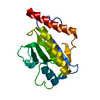
| |||||||||
|---|---|---|---|---|---|---|---|---|---|---|
| 1 |
| |||||||||
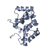
| NMR ensembles |
|
-Components
| #1: Protein | Mass: 16135.430 Da / Num. of mol.: 1 / Fragment: C-TERMINAL DOMAIN, RESIDUES 185-328 Source method: isolated from a genetically manipulated source Source: (gene. exp.) Escherichia coli (E. coli) / Plasmid: PEG041 / Production host: Escherichia coli (E. coli) / References: UniProt: P77173 |
|---|
-Experimental details
-Experiment
| Experiment | Method: SOLUTION NMR | ||||||||||||||||||||
|---|---|---|---|---|---|---|---|---|---|---|---|---|---|---|---|---|---|---|---|---|---|
| NMR experiment |
| ||||||||||||||||||||
| NMR details | Text: The structure was determined using triple-resonance NMR spectroscopy. refinement program: x-plor V3.840, authors: brunger |
- Sample preparation
| Details |
| ||||||||||||||||||||
|---|---|---|---|---|---|---|---|---|---|---|---|---|---|---|---|---|---|---|---|---|---|
| Sample conditions |
| ||||||||||||||||||||
| Crystal grow | *PLUS Method: other / Details: NMR |
-NMR measurement
| NMR spectrometer | Type: Bruker DRX / Manufacturer: Bruker / Model: DRX / Field strength: 600 MHz |
|---|
- Processing
| NMR software |
| ||||||||||||||||||||||||
|---|---|---|---|---|---|---|---|---|---|---|---|---|---|---|---|---|---|---|---|---|---|---|---|---|---|
| Refinement | Method: distance geometry simulated annealing / Software ordinal: 1 Details: The structures are based on a total of 2758 restraints, 2038 are NOE-derived distance constraints, 377 dihedral angle restraints, 84 distance restraints from hydrogen bonds, 113 3JNHa ...Details: The structures are based on a total of 2758 restraints, 2038 are NOE-derived distance constraints, 377 dihedral angle restraints, 84 distance restraints from hydrogen bonds, 113 3JNHa coupling restraints, 230 secondary Ca/Cb chemical shift restraints, and a conformational database. The coordinates in this entry corrospond to the refined minimized average structure determined from an ensemble of 30 structures | ||||||||||||||||||||||||
| NMR representative | Selection criteria: nmr, minimized average structure | ||||||||||||||||||||||||
| NMR ensemble | Conformers submitted total number: 1 |
