 Movie
Movie Controller


+ Open data

- Basic information
| Entry | Database: PDB / ID: 1ega | ||||||
|---|---|---|---|---|---|---|---|
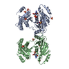
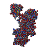
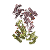
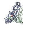
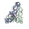
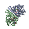
| Title | CRYSTAL STRUCTURE OF A WIDELY CONSERVED GTPASE ERA | ||||||
 Components Components | PROTEIN (GTP-BINDING PROTEIN ERA) | ||||||
 Keywords Keywords |  HYDROLASE / ERA / GTPASE / RNA-BINDING / RAS-LIKE HYDROLASE / ERA / GTPASE / RNA-BINDING / RAS-LIKE | ||||||
| Function / homology |  Function and homology informationguanosine tetraphosphate binding / ribosomal small subunit binding / ribosomal small subunit biogenesis / small ribosomal subunit rRNA binding / ribosomal small subunit assembly / rRNA binding / protein phosphorylation / GTPase activity / GTP binding / RNA binding ...guanosine tetraphosphate binding / ribosomal small subunit binding / ribosomal small subunit biogenesis / small ribosomal subunit rRNA binding / ribosomal small subunit assembly / rRNA binding / protein phosphorylation / GTPase activity / GTP binding / RNA binding / plasma membrane / cytosol / cytoplasm Function and homology informationguanosine tetraphosphate binding / ribosomal small subunit binding / ribosomal small subunit biogenesis / small ribosomal subunit rRNA binding / ribosomal small subunit assembly / rRNA binding / protein phosphorylation / GTPase activity / GTP binding / RNA binding ...guanosine tetraphosphate binding / ribosomal small subunit binding / ribosomal small subunit biogenesis / small ribosomal subunit rRNA binding / ribosomal small subunit assembly / rRNA binding / protein phosphorylation / GTPase activity / GTP binding / RNA binding / plasma membrane / cytosol / cytoplasmSimilarity search - Function | ||||||
| Biological species |  Escherichia coli (E. coli) Escherichia coli (E. coli) | ||||||
| Method | X-RAY DIFFRACTION / MIR / Resolution: 2.4 Å | ||||||
 Authors Authors | Chen, X. / Ji, X. | ||||||
 Citation Citation | Journal: Proc Natl Acad Sci U S A / Year: 1999 Title: Crystal structure of ERA: a GTPase-dependent cell cycle regulator containing an RNA binding motif. Authors: X Chen / D L Court / X Ji /  Abstract: ERA forms a unique family of GTPase. It is widely conserved and essential in bacteria. ERA functions in cell cycle control by coupling cell division with growth rate. ERA homologues also are found in ...ERA forms a unique family of GTPase. It is widely conserved and essential in bacteria. ERA functions in cell cycle control by coupling cell division with growth rate. ERA homologues also are found in eukaryotes. Here we report the crystal structure of ERA from Escherichia coli. The structure has been determined at 2.4-A resolution. It reveals a two-domain arrangement of the molecule: an N-terminal domain that resembles p21 Ras and a C-terminal domain that is unique. Structure-based topological search of the C domain fails to reveal any meaningful match, although sequence analysis suggests that it contains a KH domain. KH domains are RNA binding motifs that usually occur in tandem repeats and exhibit low sequence similarity except for the well-conserved segment VIGxxGxxIK. We have identified a betaalphaalphabeta fold that contains the VIGxxGxxIK sequence and is shared by the C domain of ERA and the KH domain. We propose that this betaalphaalphabeta fold is the RNA binding motif, the minimum structural requirement for RNA binding. ERA dimerizes in crystal. The dimer formation involves a significantly distorted switch II region, which may shed light on how ERA protein regulates downstream events. #1: Journal: Mol.Microbiol. / Year: 1998Title: Cell Cycle Arrest in Era GTPase Mutants: A Potential Growth Rate-Regulated Checkpoint in E. Coli Authors: Britton, R.A. / Powell, B.S. / Dasgupta, S. / Sun, Q. / Margolin, W. / Lupski, J.R. / Court, D.L. | ||||||
| History |
|
- Structure visualization
| Structure viewer | Molecule: MolmilJmol/JSmol |
|---|
- Downloads & links
-Download
| PDBx/mmCIF format | 1ega.cif.gz | 128.1 KB | Display | PDBx/mmCIF format |
|---|---|---|---|---|
| PDB format | pdb1ega.ent.gz | 101 KB | Display | PDB format |
| PDBx/mmJSON format | 1ega.json.gz | Tree view | PDBx/mmJSON format | |
| Others |  Other downloads Other downloads |
-Validation report
| Arichive directory | https://data.pdbj.org/pub/pdb/validation_reports/eg/1egaftp://data.pdbj.org/pub/pdb/validation_reports/eg/1ega | HTTPS FTP |
|---|
-Related structure data
| Similar structure data |
|---|

-Links
 PDBj
PDBj- Assembly
| Deposited unit | 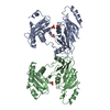
| ||||||||
|---|---|---|---|---|---|---|---|---|---|
| 1 |
| ||||||||
| Unit cell |
| ||||||||
| Noncrystallographic symmetry (NCS) | NCS oper: (Code: given Matrix: (-0.436003, 0.021836, 0.89968), Vector : |
-Components
| #1: Protein | Mass: 33858.078 Da / Num. of mol.: 2 / Source method: isolated from a natural source / Source: (natural) Escherichia coli (E. coli) / Cellular location: CYTOPLASM/MEMBRANE / Strain: TAP106 / References: UniProt: P06616#2: Chemical | ChemComp-SO4 / Sulfate  Mass: 96.063 Da / Num. of mol.: 5 / Source method: obtained synthetically / Formula: SO4 Mass: 96.063 Da / Num. of mol.: 5 / Source method: obtained synthetically / Formula: SO4#3: Water | ChemComp-HOH / | Water Mass: 18.015 Da / Num. of mol.: 149 / Source method: isolated from a natural source / Formula: H2O Mass: 18.015 Da / Num. of mol.: 149 / Source method: isolated from a natural source / Formula: H2O |
|---|
-Experimental details
-Experiment
| Experiment | Method: X-RAY DIFFRACTION / Number of used crystals: 1 |
|---|
- Sample preparation
| Crystal | Density Matthews: 3.5 Å3/Da / Density % sol: 65 % Description: THE STRUCTURE WAS SOLVED BY A COMBINATION OF MIR AND MAD METHODS. MAD DATA SETS, USING HG ANOMALOUS SCATTERING AT WAVELENGTH 1.00870A, 1.00764A, 0.99184A AND 1.00903A, WERE COLLECTED AT ...Description: THE STRUCTURE WAS SOLVED BY A COMBINATION OF MIR AND MAD METHODS. MAD DATA SETS, USING HG ANOMALOUS SCATTERING AT WAVELENGTH 1.00870A, 1.00764A, 0.99184A AND 1.00903A, WERE COLLECTED AT NSLS BEAMLINE X9B WITH MAR345 DETECTOR. | ||||||||||||||||||||||||||||||
|---|---|---|---|---|---|---|---|---|---|---|---|---|---|---|---|---|---|---|---|---|---|---|---|---|---|---|---|---|---|---|---|
| Crystal grow | pH: 8 Details: PROTEIN 8MG/ML TRIS BUFFER 0.1M, PH 8.0 LITHIUM SULFATE 0.8M SODIUM CHLORIDE 0.8M | ||||||||||||||||||||||||||||||
| Components of the solutions |
| ||||||||||||||||||||||||||||||
| Crystal grow | *PLUS Method: vapor diffusion, hanging drop | ||||||||||||||||||||||||||||||
| Components of the solutions | *PLUS
|
-Data collection
| Diffraction | Mean temperature: 100 K |
|---|---|
| Diffraction source | Source: ROTATING ANODE / Type: RIGAKU / Wavelength: 1.5418 |
| Detector | Type: MARRESEARCH / Detector: IMAGE PLATE / Date: Jun 18, 1998 / Details: BENT MIRRORS |
| Radiation | Monochromator: GRAPHITE / Protocol: SINGLE WAVELENGTH / Monochromatic (M) / Laue (L): M / Scattering type: x-ray |
| Radiation wavelength | Wavelength: 1.5418 Å / Relative weight: 1 |
| Reflection | Resolution: 2.4→20 Å / Num. obs: 35696 / % possible obs: 99.7 % / Observed criterion σ(I): 0 / Redundancy: 6.4 % / Biso Wilson estimate: 53.4 Å2 / Rsym value: 0.056 / Net I/σ(I): 31.8 |
| Reflection shell | Resolution: 2.4→2.5 Å / Redundancy: 6.1 % / Mean I/σ(I) obs: 3.9 / Rsym value: 0.49 / % possible all: 99.1 |
| Reflection | *PLUS Rmerge(I) obs: 0.056 |
| Reflection shell | *PLUS % possible obs: 99.1 % / Rmerge(I) obs: 0.49 |
- Processing
| Software |
| ||||||||||||||||||||||||||||||||||||||||||||||||||||||||||||
|---|---|---|---|---|---|---|---|---|---|---|---|---|---|---|---|---|---|---|---|---|---|---|---|---|---|---|---|---|---|---|---|---|---|---|---|---|---|---|---|---|---|---|---|---|---|---|---|---|---|---|---|---|---|---|---|---|---|---|---|---|---|
| Refinement | Method to determine structure: MIR / Resolution: 2.4→8 Å / Rfactor Rfree error: 0.0086 / Data cutoff high absF: 100000 / Data cutoff low absF: 0.1 / Cross valid method: THROUGHOUT / σ(F): 4 Details: N-TERMINAL RESIDUES 1-3 FOR BOTH CHAINS, AND C-TERMINAL RESIDUES 296-301 FOR CHAIN A AND RESIDUES 297-301 FOR CHAIN B WERE NOT OBSERVED IN THE ELECTRON DNESITY AND NOT INCLUDED IN THE MODEL; ...Details: N-TERMINAL RESIDUES 1-3 FOR BOTH CHAINS, AND C-TERMINAL RESIDUES 296-301 FOR CHAIN A AND RESIDUES 297-301 FOR CHAIN B WERE NOT OBSERVED IN THE ELECTRON DNESITY AND NOT INCLUDED IN THE MODEL; RESIDUES 224 - 228 IN BOTH CHAINS WERE NOT OBSERVED IN THE ELECTRON DENSITY AND NOT INCLUDED IN THE REFINEMENT, BUT BUILT STEREOCHEMICALLY AND INCLUDED IN THE MODEL; RESTRAINTS BETWEEN THE TWO COPIES OF MOLECULES WERE APPLIED DURING THE INITIAL STAGE OF THE REFINEMENT ONLY
| ||||||||||||||||||||||||||||||||||||||||||||||||||||||||||||
| Displacement parameters | Biso mean: 49.5 Å2 | ||||||||||||||||||||||||||||||||||||||||||||||||||||||||||||
| Refinement step | Cycle: LAST / Resolution: 2.4→8 Å
| ||||||||||||||||||||||||||||||||||||||||||||||||||||||||||||
| Refine LS restraints |
| ||||||||||||||||||||||||||||||||||||||||||||||||||||||||||||
| LS refinement shell | Resolution: 2.4→2.5 Å / Rfactor Rfree error: 0.035 / Total num. of bins used: 8
| ||||||||||||||||||||||||||||||||||||||||||||||||||||||||||||
| Software | *PLUS Name: X-PLOR / Version: 3.85 / Classification: refinement | ||||||||||||||||||||||||||||||||||||||||||||||||||||||||||||
| Refine LS restraints | *PLUS
|
