 Movie
Movie Controller

[English] 日本語
 Yorodumi
- PDB-3lou: Crystal structure of Formyltetrahydrofolate deformylase (YP_10525... -
+ Open data

- Basic information
| Entry | Database: PDB / ID: 3lou | ||||||
|---|---|---|---|---|---|---|---|
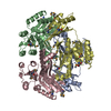
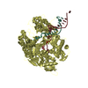
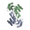
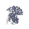
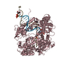
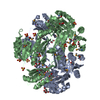
| Title | Crystal structure of Formyltetrahydrofolate deformylase (YP_105254.1) from BURKHOLDERIA MALLEI ATCC 23344 at 1.90 A resolution | ||||||
 Components Components | Formyltetrahydrofolate deformylase | ||||||
 Keywords Keywords | HYDROLASE / Formyltetrahydrofolate deformylase / Structural Genomics / Joint Center for Structural Genomics / JCSG / Protein Structure Initiative / PSI-2 | ||||||
| Function / homology |  Function and homology informationformyltetrahydrofolate deformylase / formyltetrahydrofolate deformylase activity / hydroxymethyl-, formyl- and related transferase activity / 'de novo' IMP biosynthetic process / one-carbon metabolic process Function and homology informationformyltetrahydrofolate deformylase / formyltetrahydrofolate deformylase activity / hydroxymethyl-, formyl- and related transferase activity / 'de novo' IMP biosynthetic process / one-carbon metabolic processSimilarity search - Function | ||||||
| Biological species |  Burkholderia mallei (bacteria) Burkholderia mallei (bacteria) | ||||||
| Method | X-RAY DIFFRACTION / SYNCHROTRON / MAD / Resolution: 1.9 Å | ||||||
 Authors Authors | Joint Center for Structural Genomics (JCSG) | ||||||
 Citation Citation | Journal: To be published Title: Crystal structure of Formyltetrahydrofolate deformylase (YP_105254.1) from BURKHOLDERIA MALLEI ATCC 23344 at 1.90 A resolution Authors: Joint Center for Structural Genomics (JCSG) | ||||||
| History |
|
- Structure visualization
| Structure viewer | Molecule: MolmilJmol/JSmol |
|---|
- Downloads & links
-Download
| PDBx/mmCIF format | 3lou.cif.gz | 259 KB | Display | PDBx/mmCIF format |
|---|---|---|---|---|
| PDB format | pdb3lou.ent.gz | 213.8 KB | Display | PDB format |
| PDBx/mmJSON format | 3lou.json.gz | Tree view | PDBx/mmJSON format | |
| Others |  Other downloads Other downloads |
-Validation report
| Arichive directory | https://data.pdbj.org/pub/pdb/validation_reports/lo/3louftp://data.pdbj.org/pub/pdb/validation_reports/lo/3lou | HTTPS FTP |
|---|
-Related structure data
| Similar structure data | |
|---|---|
| Other databases |
-Links
 PDBj
PDBj

- Assembly
| Deposited unit | 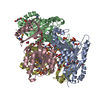
| |||||||||||||||||||||||||||||||||||||||||||||
|---|---|---|---|---|---|---|---|---|---|---|---|---|---|---|---|---|---|---|---|---|---|---|---|---|---|---|---|---|---|---|---|---|---|---|---|---|---|---|---|---|---|---|---|---|---|---|
| 1 |
| |||||||||||||||||||||||||||||||||||||||||||||
| 2 | 
| |||||||||||||||||||||||||||||||||||||||||||||
| 3 | 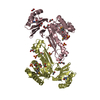
| |||||||||||||||||||||||||||||||||||||||||||||
| Unit cell |
| |||||||||||||||||||||||||||||||||||||||||||||
| Noncrystallographic symmetry (NCS) | NCS domain:
NCS domain segments:
| |||||||||||||||||||||||||||||||||||||||||||||
| Details | ANALYTICAL SIZE EXCLUSION CHROMATOGRAPHY SUPPORTS THE ASSIGNMENT OF A TETRAMER AS A SIGNIFICANT OLIGOMERIZATION STATE IN SOLUTION. |
-Components
-Protein , 1 types, 4 molecules ABCD
| #1: Protein | Mass: 33179.203 Da / Num. of mol.: 4 Source method: isolated from a genetically manipulated source Source: (gene. exp.) Burkholderia mallei (bacteria) / Strain: ATCC 23344 / Gene: purU-2, BMAA0482 / Plasmid: SpeedET / Production host: Escherichia Coli (E. coli) / Strain (production host): HK100References: UniProt: Q62DH4, UniProt: A0A0H2WCF8*PLUS, formyltetrahydrofolate deformylase |
|---|
-Non-polymers , 5 types, 912 molecules 


| #2: Chemical | ChemComp-UNL / Num. of mol.: 1 / Source method: obtained synthetically | ||||||
|---|---|---|---|---|---|---|---|
| #3: Chemical | ChemComp-SO4 / Sulfate Mass: 96.063 Da / Num. of mol.: 12 / Source method: obtained synthetically / Formula: SO4 Mass: 96.063 Da / Num. of mol.: 12 / Source method: obtained synthetically / Formula: SO4#4: Chemical | ChemComp-EDO / Ethylene glycol Mass: 62.068 Da / Num. of mol.: 23 / Source method: obtained synthetically / Formula: C2H6O2 Mass: 62.068 Da / Num. of mol.: 23 / Source method: obtained synthetically / Formula: C2H6O2#5: Chemical | ChemComp-PEG / Diethylene glycol Mass: 106.120 Da / Num. of mol.: 7 / Source method: obtained synthetically / Formula: C4H10O3 Mass: 106.120 Da / Num. of mol.: 7 / Source method: obtained synthetically / Formula: C4H10O3#6: Water | ChemComp-HOH / | WaterMass: 18.015 Da / Num. of mol.: 869 / Source method: isolated from a natural source / Formula: H2O |
-Details
| Sequence details | SEQUENCE THIS CONSTRUCT WAS EXPRESSED WITH A PURIFICATION TAG MGSDKIHHHHHHENLYFQG. THE TAG WAS ...SEQUENCE THIS CONSTRUCT WAS EXPRESSED WITH A PURIFICATI |
|---|
-Experimental details
-Experiment
| Experiment | Method: X-RAY DIFFRACTION / Number of used crystals: 1 |
|---|
- Sample preparation
| Crystal | Density Matthews: 2.59 Å3/Da / Density % sol: 52.43 % |
|---|---|
| Crystal grow | Temperature: 277 K / Method: vapor diffusion, sitting drop / pH: 8 Details: 0.2000M Li2SO4, 10.0000% PEG-3000, 0.1M Imidazole pH 8.0, NANODROP, VAPOR DIFFUSION, SITTING DROP, temperature 277K |
-Data collection
| Diffraction | Mean temperature: 100 K | |||||||||||||||||||||||||||||||||||||||||||||||||||||||||||||||||||||||||||||
|---|---|---|---|---|---|---|---|---|---|---|---|---|---|---|---|---|---|---|---|---|---|---|---|---|---|---|---|---|---|---|---|---|---|---|---|---|---|---|---|---|---|---|---|---|---|---|---|---|---|---|---|---|---|---|---|---|---|---|---|---|---|---|---|---|---|---|---|---|---|---|---|---|---|---|---|---|---|---|
| Diffraction source | Source: SYNCHROTRON / Site: SSRL  / Beamline: BL9-2 / Wavelength: 0.91837,0.97922 / Beamline: BL9-2 / Wavelength: 0.91837,0.97922 | |||||||||||||||||||||||||||||||||||||||||||||||||||||||||||||||||||||||||||||
| Detector | Type: MARMOSAIC 325 mm CCD / Detector: CCD / Date: Jul 31, 2009 / Details: Flat collimating mirror, toroid focusing mirror | |||||||||||||||||||||||||||||||||||||||||||||||||||||||||||||||||||||||||||||
| Radiation | Monochromator: Double crystal monochromator / Protocol: MAD / Monochromatic (M) / Laue (L): M / Scattering type: x-ray | |||||||||||||||||||||||||||||||||||||||||||||||||||||||||||||||||||||||||||||
| Radiation wavelength |
| |||||||||||||||||||||||||||||||||||||||||||||||||||||||||||||||||||||||||||||
| Reflection | Resolution: 1.9→29.488 Å / Num. obs: 105637 / % possible obs: 98 % / Observed criterion σ(I): -3 / Biso Wilson estimate: 25.053 Å2 / Rmerge(I) obs: 0.069 / Net I/σ(I): 7.73 | |||||||||||||||||||||||||||||||||||||||||||||||||||||||||||||||||||||||||||||
| Reflection shell |
|
-Phasing
| Phasing | Method: MAD |
|---|
- Processing
| Software |
| |||||||||||||||||||||||||||||||||||||||||||||||||||||||||||||||||||||||||||||||||||||||||||||||||||||||||||||||||||||||||||||
|---|---|---|---|---|---|---|---|---|---|---|---|---|---|---|---|---|---|---|---|---|---|---|---|---|---|---|---|---|---|---|---|---|---|---|---|---|---|---|---|---|---|---|---|---|---|---|---|---|---|---|---|---|---|---|---|---|---|---|---|---|---|---|---|---|---|---|---|---|---|---|---|---|---|---|---|---|---|---|---|---|---|---|---|---|---|---|---|---|---|---|---|---|---|---|---|---|---|---|---|---|---|---|---|---|---|---|---|---|---|---|---|---|---|---|---|---|---|---|---|---|---|---|---|---|---|---|
| Refinement | Method to determine structure: MAD / Resolution: 1.9→29.488 Å / Cor.coef. Fo:Fc: 0.955 / Cor.coef. Fo:Fc free: 0.941 / Occupancy max: 1 / Occupancy min: 0.33 / SU B: 7.289 / SU ML: 0.097 / TLS residual ADP flag: LIKELY RESIDUAL / Cross valid method: THROUGHOUT / σ(F): 0 / ESU R: 0.15 / ESU R Free: 0.137 Stereochemistry target values: MAXIMUM LIKELIHOOD WITH PHASES Details: 1.HYDROGENS HAVE BEEN ADDED IN THE RIDING POSITIONS. 2.ATOM RECORDS CONTAIN RESIDUAL B FACTORS ONLY. 3.A MET-INHIBITION PROTOCOL WAS USED FOR SELENOMETHIONINE INCORPORATION DURING PROTEIN ...Details: 1.HYDROGENS HAVE BEEN ADDED IN THE RIDING POSITIONS. 2.ATOM RECORDS CONTAIN RESIDUAL B FACTORS ONLY. 3.A MET-INHIBITION PROTOCOL WAS USED FOR SELENOMETHIONINE INCORPORATION DURING PROTEIN EXPRESSION. THE OCCUPANCY OF THE SE ATOMS IN THE MSE RESIDUES WAS REDUCED TO 0.75 FOR THE REDUCED SCATTERING POWER DUE TO PARTIAL S-MET INCORPORATION. 4. AN UNKNOWN LIGAND (UNL) HAS BEEN MODELED INTO THE STRUCTURE. 5. SULFATE (SO4),POLYETHYLENE GLYCOL, AND ETHYLENE GLYCOL FROM THE CRYSTALLIZATION/CRYOGENIC CONDITIONS HAVE BEEN MODELED INTO THE STRUCTURE. 6. ELECTRON DENSITIES BETWEEN RESIDUES 62-65 ON THE A AND C SUBUNITS, AND 61-65 ON THE B AND D SUBUNITS ARE DISORDERED; THEREFORE, THESE RESIDUES WERE NOT MODELED. BULK SOLVENT MODELLING. METHOD USED : BABINET MODEL WITH MASK PARAMETERS FOR MASK CALCULATION VDW PROBE RADIUS : 1.40 ION PROBE RADIUS : 0.80 SHRINKAGE RADIUS : 0.80
| |||||||||||||||||||||||||||||||||||||||||||||||||||||||||||||||||||||||||||||||||||||||||||||||||||||||||||||||||||||||||||||
| Displacement parameters | Biso max: 75.91 Å2 / Biso mean: 26.272 Å2 / Biso min: 10.4 Å2
| |||||||||||||||||||||||||||||||||||||||||||||||||||||||||||||||||||||||||||||||||||||||||||||||||||||||||||||||||||||||||||||
| Refinement step | Cycle: LAST / Resolution: 1.9→29.488 Å
| |||||||||||||||||||||||||||||||||||||||||||||||||||||||||||||||||||||||||||||||||||||||||||||||||||||||||||||||||||||||||||||
| Refine LS restraints |
| |||||||||||||||||||||||||||||||||||||||||||||||||||||||||||||||||||||||||||||||||||||||||||||||||||||||||||||||||||||||||||||
| Refine LS restraints NCS | Ens-ID: 1 / Number: 3297 / Refine-ID: X-RAY DIFFRACTION
| |||||||||||||||||||||||||||||||||||||||||||||||||||||||||||||||||||||||||||||||||||||||||||||||||||||||||||||||||||||||||||||
| LS refinement shell | Resolution: 1.9→1.95 Å / Total num. of bins used: 20
| |||||||||||||||||||||||||||||||||||||||||||||||||||||||||||||||||||||||||||||||||||||||||||||||||||||||||||||||||||||||||||||
| Refinement TLS params. | Method: refined / Refine-ID: X-RAY DIFFRACTION
| |||||||||||||||||||||||||||||||||||||||||||||||||||||||||||||||||||||||||||||||||||||||||||||||||||||||||||||||||||||||||||||
| Refinement TLS group |
|
