 Movie
Movie Controller

[English] 日本語
 Yorodumi
- PDB-1dev: CRYSTAL STRUCTURE OF SMAD2 MH2 DOMAIN BOUND TO THE SMAD-BINDING D... -
+ Open data

- Basic information
| Entry | Database: PDB / ID: 1dev | ||||||
|---|---|---|---|---|---|---|---|
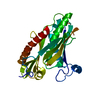
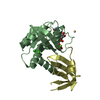
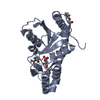
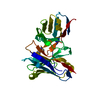
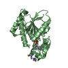
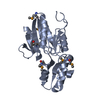
| Title | CRYSTAL STRUCTURE OF SMAD2 MH2 DOMAIN BOUND TO THE SMAD-BINDING DOMAIN OF SARA | ||||||
 Components Components |
| ||||||
 Keywords Keywords |  SIGNALING PROTEIN / BETA SHEET / THREE-HELIX BUNDLE SIGNALING PROTEIN / BETA SHEET / THREE-HELIX BUNDLE | ||||||
| Function / homology |  Function and homology information Function and homology informationzygotic specification of dorsal/ventral axis / regulation of binding / homomeric SMAD protein complex / paraxial mesoderm morphogenesis / activin responsive factor complex / SMAD4 MH2 Domain Mutants in Cancer / SMAD2/3 MH2 Domain Mutants in Cancer / nodal signaling pathway / SMAD protein complex / 1-phosphatidylinositol binding ...zygotic specification of dorsal/ventral axis / regulation of binding / homomeric SMAD protein complex / paraxial mesoderm morphogenesis / activin responsive factor complex / SMAD4 MH2 Domain Mutants in Cancer / SMAD2/3 MH2 Domain Mutants in Cancer / nodal signaling pathway / SMAD protein complex / 1-phosphatidylinositol binding / endoderm formation / co-SMAD binding / heteromeric SMAD protein complex / determination of left/right asymmetry in lateral mesoderm / odontoblast differentiation / secondary palate development / pericardium development / FOXO-mediated transcription of cell cycle genes / SMAD2/3 Phosphorylation Motif Mutants in Cancer / TGFBR1 KD Mutants in Cancer / Transcriptional regulation of pluripotent stem cells / regulation of transforming growth factor beta receptor signaling pathway / embryonic foregut morphogenesis / transforming growth factor beta receptor binding / primary miRNA processing / Germ layer formation at gastrulation / pulmonary valve morphogenesis / type I transforming growth factor beta receptor binding / Formation of definitive endoderm / activin receptor signaling pathway / signal transduction involved in regulation of gene expression / Signaling by Activin / SMAD protein signal transduction / Formation of axial mesoderm / positive regulation of BMP signaling pathway / Signaling by NODAL / embryonic cranial skeleton morphogenesis / response to cholesterol / I-SMAD binding / aortic valve morphogenesis / pancreas development / insulin secretion / anterior/posterior pattern specification / ureteric bud development / endocardial cushion morphogenesis / endosomal transport / organ growth / adrenal gland development / SMAD binding / TGF-beta receptor signaling activates SMADs / R-SMAD binding / mesoderm formation / anatomical structure morphogenesis / phosphatase binding / cell fate commitment / cis-regulatory region sequence-specific DNA binding / FOXO-mediated transcription of oxidative stress, metabolic and neuronal genes / positive regulation of epithelial to mesenchymal transition / gastrulation / Downregulation of TGF-beta receptor signaling / transforming growth factor beta receptor signaling pathway / post-embryonic development / cellular response to glucose stimulus / Downregulation of SMAD2/3:SMAD4 transcriptional activity / SMAD2/SMAD3:SMAD4 heterotrimer regulates transcription / lung development / tau protein binding / endocytosis / disordered domain specific binding / early endosome membrane / double-stranded DNA binding / DNA-binding transcription activator activity, RNA polymerase II-specific / DNA-binding transcription factor binding / in utero embryonic development / RNA polymerase II-specific DNA-binding transcription factor binding / cell population proliferation / transcription regulator complex / early endosome / cell differentiation / DNA-binding transcription factor activity, RNA polymerase II-specific / Ub-specific processing proteases / intracellular signal transduction / RNA polymerase II cis-regulatory region sequence-specific DNA binding / DNA-binding transcription factor activity / protein domain specific binding / negative regulation of cell population proliferation / intracellular membrane-bounded organelle / negative regulation of DNA-templated transcription / DNA-templated transcription / ubiquitin protein ligase binding / chromatin binding / chromatin / regulation of DNA-templated transcription / positive regulation of gene expression / positive regulation of DNA-templated transcription / positive regulation of transcription by RNA polymerase II / protein-containing complex / nucleoplasm / identical protein binding / metal ion bindingSimilarity search - Function | ||||||
| Biological species |  Homo sapiens (human) Homo sapiens (human) | ||||||
| Method | X-RAY DIFFRACTION / SYNCHROTRON / Resolution: 2.2 Å | ||||||
 Authors Authors | Shi, Y. / Wu, G. | ||||||
 Citation Citation | Journal: Science / Year: 2000 Title: Structural basis of Smad2 recognition by the Smad anchor for receptor activation. Authors: Wu, G. / Chen, Y.G. / Ozdamar, B. / Gyuricza, C.A. / Chong, P.A. / Wrana, J.L. / Massague, J. / Shi, Y. | ||||||
| History |
|
- Structure visualization
| Structure viewer | Molecule: MolmilJmol/JSmol |
|---|
- Downloads & links
-Download
| PDBx/mmCIF format | 1dev.cif.gz | 99.7 KB | Display | PDBx/mmCIF format |
|---|---|---|---|---|
| PDB format | pdb1dev.ent.gz | 78.2 KB | Display | PDB format |
| PDBx/mmJSON format | 1dev.json.gz | Tree view | PDBx/mmJSON format | |
| Others |  Other downloads Other downloads |
-Validation report
| Arichive directory | https://data.pdbj.org/pub/pdb/validation_reports/de/1devftp://data.pdbj.org/pub/pdb/validation_reports/de/1dev | HTTPS FTP |
|---|
-Related structure data
| Similar structure data |
|---|


-Links
 PDBj
PDBj


- Assembly
| Deposited unit | 
| ||||||||
|---|---|---|---|---|---|---|---|---|---|
| 1 | 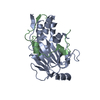
| ||||||||
| 2 | 
| ||||||||
| Unit cell |
| ||||||||
| Details | The biological assembly is a hetero-dimer of Smad2 and SARA. |
-Components
| #1: Protein | Mass: 22276.291 Da / Num. of mol.: 2 / Fragment: SMAD2 MH2 DOMAIN Source method: isolated from a genetically manipulated source Source: (gene. exp.) Homo sapiens (human) / Description: THIS SEQUENCE OCCURS NATURALLY IN HUMANS / Plasmid: PET3 AND PGEX / Production host:  Escherichia coli (E. coli) / References: UniProt: Q15796 Escherichia coli (E. coli) / References: UniProt: Q15796#2: Protein/peptide | Zinc finger FYVE domain-containing protein 9Mass: 4132.583 Da / Num. of mol.: 2 / Fragment: SARA SMAD2-BINDING DOMAIN Source method: isolated from a genetically manipulated source Source: (gene. exp.) Homo sapiens (human) / Description: THIS SEQUENCE OCCURS NATURALLY IN HUMANS / Plasmid: PET3 AND PGEX / Production host: Escherichia coli (E. coli) / References: UniProt: O95405 |
|---|
-Experimental details
-Experiment
| Experiment | Method: X-RAY DIFFRACTION / Number of used crystals: 1 |
|---|
- Sample preparation
| Crystal | Density Matthews: 2.93 Å3/Da / Density % sol: 57.99 % | |||||||||||||||||||||||||
|---|---|---|---|---|---|---|---|---|---|---|---|---|---|---|---|---|---|---|---|---|---|---|---|---|---|---|
| Crystal grow | Temperature: 277 K / Method: vapor diffusion, hanging drop / pH: 8 Details: TRIS, DIOXANE, AMMONIUM SULFATE, pH 8.0, VAPOR DIFFUSION, HANGING DROP, temperature 4K | |||||||||||||||||||||||||
| Crystal grow | *PLUS pH: 8.5 | |||||||||||||||||||||||||
| Components of the solutions | *PLUS
|
-Data collection
| Diffraction | Mean temperature: 100 K |
|---|---|
| Diffraction source | Source: SYNCHROTRON / Site: NSLS  / Beamline: X25 / Wavelength: 1.15 / Beamline: X25 / Wavelength: 1.15 |
| Detector | Type: FUJI / Detector: IMAGE PLATE / Date: Sep 1, 1999 |
| Radiation | Protocol: SINGLE WAVELENGTH / Monochromatic (M) / Laue (L): M / Scattering type: x-ray |
| Radiation wavelength | Wavelength: 1.15 Å / Relative weight: 1 |
| Reflection | Resolution: 2.2→20 Å / Num. all: 31596 / Num. obs: 31296 / % possible obs: 100 % / Observed criterion σ(F): 2 / Observed criterion σ(I): 2 / Redundancy: 9 % / Biso Wilson estimate: 37 Å2 / Rmerge(I) obs: 0.039 / Net I/σ(I): 45 |
| Reflection shell | Resolution: 2.2→2.28 Å / Redundancy: 3 % / Rmerge(I) obs: 0.167 / % possible all: 97.6 |
| Reflection | *PLUS Num. measured all: 218815 |
| Reflection shell | *PLUS % possible obs: 97.6 % |
- Processing
| Software |
| ||||||||||||||||||||||||||||||||||||||||||||||||||||||||||||
|---|---|---|---|---|---|---|---|---|---|---|---|---|---|---|---|---|---|---|---|---|---|---|---|---|---|---|---|---|---|---|---|---|---|---|---|---|---|---|---|---|---|---|---|---|---|---|---|---|---|---|---|---|---|---|---|---|---|---|---|---|---|
| Refinement | Resolution: 2.2→20 Å / σ(F): 1.414 / σ(I): 2 / Stereochemistry target values: ENGH & HUBER
| ||||||||||||||||||||||||||||||||||||||||||||||||||||||||||||
| Refinement step | Cycle: LAST / Resolution: 2.2→20 Å
| ||||||||||||||||||||||||||||||||||||||||||||||||||||||||||||
| Refine LS restraints |
| ||||||||||||||||||||||||||||||||||||||||||||||||||||||||||||
| Software | *PLUS Name: X-PLOR / Version: 3.851 / Classification: refinement | ||||||||||||||||||||||||||||||||||||||||||||||||||||||||||||
| Refinement | *PLUS | ||||||||||||||||||||||||||||||||||||||||||||||||||||||||||||
| Solvent computation | *PLUS | ||||||||||||||||||||||||||||||||||||||||||||||||||||||||||||
| Displacement parameters | *PLUS |
