 Movie
Movie Controller


+ Open data

- Basic information
| Entry | Database: PDB / ID: 1ddw | ||||||
|---|---|---|---|---|---|---|---|
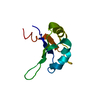
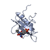
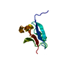
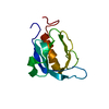
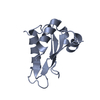
| Title | HOMER EVH1 DOMAIN UNLIGANDED | ||||||
 Components Components | GLGF-DOMAIN PROTEIN HOMER | ||||||
 Keywords Keywords |  SIGNALING PROTEIN / PLECKSTRIN HOMOLOGY DOMAIN FOLD SIGNALING PROTEIN / PLECKSTRIN HOMOLOGY DOMAIN FOLD | ||||||
| Function / homology |  Function and homology information Function and homology informationG protein-coupled glutamate receptor binding / structural constituent of postsynapse / regulation of dendritic spine maintenance / regulation of store-operated calcium entry / Neurexins and neuroligins / regulation of calcium ion import / G protein-coupled glutamate receptor signaling pathway / protein localization to synapse / neuron spine / costamere ...G protein-coupled glutamate receptor binding / structural constituent of postsynapse / regulation of dendritic spine maintenance / regulation of store-operated calcium entry / Neurexins and neuroligins / regulation of calcium ion import / G protein-coupled glutamate receptor signaling pathway / protein localization to synapse / neuron spine / costamere / positive regulation of calcium ion transport / type 5 metabotropic glutamate receptor binding / postsynaptic cytosol / behavioral response to cocaine / skeletal muscle contraction / skeletal muscle fiber development / regulation of synaptic transmission, glutamatergic / dendritic shaft / response to nicotine / response to cocaine / protein tetramerization / Z disc / response to calcium ion / circadian rhythm / apical part of cell / scaffold protein binding / postsynapse / transmembrane transporter binding / dendritic spine / postsynaptic density / molecular adaptor activity / neuron projection / axon / signaling receptor binding / neuronal cell body / glutamatergic synapse / dendrite / protein-containing complex binding / membrane / identical protein binding / plasma membrane / cytosol / cytoplasmSimilarity search - Function | ||||||
| Biological species |  Rattus norvegicus (Norway rat) Rattus norvegicus (Norway rat) | ||||||
| Method | X-RAY DIFFRACTION / SYNCHROTRON / Resolution: 1.7 Å | ||||||
 Authors Authors | Beneken, J. / Tu, J.C. / Xiao, B. / Worley, P.F. / Leahy, D.J. | ||||||
 Citation Citation | Journal: Neuron / Year: 2000 Title: Structure of the Homer EVH1 domain-peptide complex reveals a new twist in polyproline recognition. Authors: Beneken, J. / Tu, J.C. / Xiao, B. / Nuriya, M. / Yuan, J.P. / Worley, P.F. / Leahy, D.J. #1: Journal: Neuron / Year: 1998Title: Homer binds a novel proline-rich motif and links group 1 metabotropic glutamate receptors with IP3 receptors Authors: Tu, J.C. / Xiao, B. / Yuan, J.P. / Lanahan, A.A. / Worley, P.F. / Leoffert, K. / Li, M. / Linden, D.J. | ||||||
| History |
|
- Structure visualization
| Structure viewer | Molecule: MolmilJmol/JSmol |
|---|
- Downloads & links
-Download
| PDBx/mmCIF format | 1ddw.cif.gz | 32.1 KB | Display | PDBx/mmCIF format |
|---|---|---|---|---|
| PDB format | pdb1ddw.ent.gz | 24.8 KB | Display | PDB format |
| PDBx/mmJSON format | 1ddw.json.gz | Tree view | PDBx/mmJSON format | |
| Others |  Other downloads Other downloads |
-Validation report
| Arichive directory | https://data.pdbj.org/pub/pdb/validation_reports/dd/1ddwftp://data.pdbj.org/pub/pdb/validation_reports/dd/1ddw | HTTPS FTP |
|---|
-Related structure data


-Links
 PDBj
PDBj- Assembly
| Deposited unit | 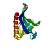
| ||||||||
|---|---|---|---|---|---|---|---|---|---|
| 1 |
| ||||||||
| Unit cell |
|
-Components
| #1: Protein | Mass: 13827.220 Da / Num. of mol.: 1 / Fragment: HOMER EVH1 DOMAIN RESIDUES 1-120 / Mutation: NONE Source method: isolated from a genetically manipulated source Source: (gene. exp.) Rattus norvegicus (Norway rat) / Organ: BRAIN / Plasmid: PGEX / Production host: Bacteria (eubacteria) / References: UniProt: Q9Z214 |
|---|---|
| #2: Water | ChemComp-HOH / Water Mass: 18.015 Da / Num. of mol.: 88 / Source method: isolated from a natural source / Formula: H2O Mass: 18.015 Da / Num. of mol.: 88 / Source method: isolated from a natural source / Formula: H2O |
-Experimental details
-Experiment
| Experiment | Method: X-RAY DIFFRACTION / Number of used crystals: 1 |
|---|
- Sample preparation
| Crystal | Density Matthews: 2.11 Å3/Da / Density % sol: 41.59 % | ||||||||||||||||||||||||||||||
|---|---|---|---|---|---|---|---|---|---|---|---|---|---|---|---|---|---|---|---|---|---|---|---|---|---|---|---|---|---|---|---|
| Crystal grow | Temperature: 293 K / Method: vapor diffusion, hanging drop / pH: 7.4 Details: PEG 3350, Magnesium sulfate, Hepes, pH 7.4, VAPOR DIFFUSION, HANGING DROP, temperature 293K | ||||||||||||||||||||||||||||||
| Crystal grow | *PLUS pH: 7.3 | ||||||||||||||||||||||||||||||
| Components of the solutions | *PLUS
|
-Data collection
| Diffraction |
| |||||||||||||||||||||||||
|---|---|---|---|---|---|---|---|---|---|---|---|---|---|---|---|---|---|---|---|---|---|---|---|---|---|---|
| Diffraction source |
| |||||||||||||||||||||||||
| Detector |
| |||||||||||||||||||||||||
| Radiation | Protocol: MAD / Monochromatic (M) / Laue (L): M / Scattering type: x-ray | |||||||||||||||||||||||||
| Radiation wavelength |
| |||||||||||||||||||||||||
| Reflection | Resolution: 1.7→30 Å / Num. all: 13342 / Num. obs: 12725 / % possible obs: 99.9 % / Observed criterion σ(F): 0 / Observed criterion σ(I): 0 / Redundancy: 4.8 % / Rmerge(I) obs: 0.087 / Net I/σ(I): 21 | |||||||||||||||||||||||||
| Reflection shell | Resolution: 1.7→1.78 Å / Redundancy: 2.34 % / Rmerge(I) obs: 0.286 / % possible all: 77 | |||||||||||||||||||||||||
| Reflection | *PLUS Num. obs: 24051 / % possible obs: 96.8 % / Rmerge(I) obs: 0.081 | |||||||||||||||||||||||||
| Reflection shell | *PLUS Mean I/σ(I) obs: 2.3 |

- Processing
| Software |
| ||||||||||||||||||||
|---|---|---|---|---|---|---|---|---|---|---|---|---|---|---|---|---|---|---|---|---|---|
| Refinement | Resolution: 1.7→30 Å / σ(F): 0 / σ(I): 0 / Stereochemistry target values: ENGH AND HUBER
| ||||||||||||||||||||
| Refinement step | Cycle: LAST / Resolution: 1.7→30 Å
| ||||||||||||||||||||
| Refine LS restraints |
| ||||||||||||||||||||
| Software | *PLUS Name: CNS / Classification: refinement | ||||||||||||||||||||
| Refinement | *PLUS % reflection Rfree: 10 % | ||||||||||||||||||||
| Solvent computation | *PLUS | ||||||||||||||||||||
| Displacement parameters | *PLUS |
