 Movie
Movie Controller


+ Open data

- Basic information
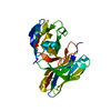
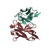
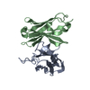
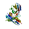
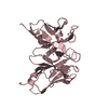
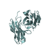
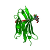
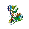
| Entry | Database: PDB / ID: 1bww | ||||||
|---|---|---|---|---|---|---|---|
| Title | BENCE-JONES IMMUNOGLOBULIN REI VARIABLE PORTION, T39K MUTANT | ||||||
 Components Components | PROTEIN (IG KAPPA CHAIN V-I REGION REI) | ||||||
 Keywords Keywords |  IMMUNE SYSTEM / REIV / STABILIZED IMMUNOGLOBULIN FRAGMENT / BENCE-JONES PROTEIN IMMUNE SYSTEM / REIV / STABILIZED IMMUNOGLOBULIN FRAGMENT / BENCE-JONES PROTEIN | ||||||
| Function / homology |  Function and homology information Function and homology informationCD22 mediated BCR regulation / Fc epsilon receptor (FCERI) signaling / Classical antibody-mediated complement activation / Initial triggering of complement / immunoglobulin complex / FCGR activation / Role of phospholipids in phagocytosis / Role of LAT2/NTAL/LAB on calcium mobilization / Scavenging of heme from plasma / antigen binding ...CD22 mediated BCR regulation / Fc epsilon receptor (FCERI) signaling / Classical antibody-mediated complement activation / Initial triggering of complement / immunoglobulin complex / FCGR activation / Role of phospholipids in phagocytosis / Role of LAT2/NTAL/LAB on calcium mobilization / Scavenging of heme from plasma / antigen binding / FCERI mediated Ca+2 mobilization / FCGR3A-mediated IL10 synthesis / Antigen activates B Cell Receptor (BCR) leading to generation of second messengers / Regulation of Complement cascade / Cell surface interactions at the vascular wall / FCERI mediated MAPK activation / FCGR3A-mediated phagocytosis / Regulation of actin dynamics for phagocytic cup formation / FCERI mediated NF-kB activation / Immunoregulatory interactions between a Lymphoid and a non-Lymphoid cell / blood microparticle / Potential therapeutics for SARS / adaptive immune response / immune response / extracellular space / extracellular exosome / extracellular region / plasma membraneSimilarity search - Function | ||||||
| Biological species |  Homo sapiens (human) Homo sapiens (human) | ||||||
| Method | X-RAY DIFFRACTION / SYNCHROTRON / MOLECULAR REPLACEMENT / Resolution: 1.7 Å | ||||||
 Authors Authors | Uson, I. / Pohl, E. / Schneider, T.R. / Dauter, Z. / Schmidt, A. / Fritz, H.J. / Sheldrick, G.M. | ||||||
 Citation Citation | Journal: Acta Crystallogr.,Sect.D / Year: 1999 Title: 1.7 A structure of the stabilized REIv mutant T39K. Application of local NCS restraints. Authors: Uson, I. / Pohl, E. / Schneider, T.R. / Dauter, Z. / Schmidt, A. / Fritz, H.J. / Sheldrick, G.M. | ||||||
| History |
|
- Structure visualization
| Structure viewer | Molecule: MolmilJmol/JSmol |
|---|
- Downloads & links
-Download
| PDBx/mmCIF format | 1bww.cif.gz | 56.3 KB | Display | PDBx/mmCIF format |
|---|---|---|---|---|
| PDB format | pdb1bww.ent.gz | 40.1 KB | Display | PDB format |
| PDBx/mmJSON format | 1bww.json.gz | Tree view | PDBx/mmJSON format | |
| Others |  Other downloads Other downloads |
-Validation report
| Arichive directory | https://data.pdbj.org/pub/pdb/validation_reports/bw/1bwwftp://data.pdbj.org/pub/pdb/validation_reports/bw/1bww | HTTPS FTP |
|---|
-Related structure data
| Related structure data | 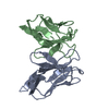 1reiS S: Starting model for refinement |
|---|---|
| Similar structure data |

-Links
 PDBj
PDBj


- Assembly
| Deposited unit | 
| ||||||||
|---|---|---|---|---|---|---|---|---|---|
| 1 |
| ||||||||
| Unit cell |
| ||||||||
| Noncrystallographic symmetry (NCS) | NCS oper: (Code: given Matrix: (-0.99977, -0.01789, 0.01212), Vector : |
-Components
| #1: Antibody | Mass: 11978.282 Da / Num. of mol.: 2 / Fragment: IMMUNOGLOBULIN KAPPA LIGHT CHAIN / Mutation: T39K Source method: isolated from a genetically manipulated source Source: (gene. exp.) Homo sapiens (human) / Cellular location (production host): PERIPLASM / Production host:  Escherichia coli (E. coli) / Keywords: ANTIBODY / References: UniProt: P01607, UniProt: P01593*PLUS Escherichia coli (E. coli) / Keywords: ANTIBODY / References: UniProt: P01607, UniProt: P01593*PLUS#2: Water | ChemComp-HOH / | Water Mass: 18.015 Da / Num. of mol.: 92 / Source method: isolated from a natural source / Formula: H2O Mass: 18.015 Da / Num. of mol.: 92 / Source method: isolated from a natural source / Formula: H2O |
|---|
-Experimental details
-Experiment
| Experiment | Method: X-RAY DIFFRACTION / Number of used crystals: 1 |
|---|
- Sample preparation
| Crystal | Density Matthews: 2.26 Å3/Da / Density % sol: 45 % | |||||||||||||||||||||||||
|---|---|---|---|---|---|---|---|---|---|---|---|---|---|---|---|---|---|---|---|---|---|---|---|---|---|---|
| Crystal grow | pH: 7 Details: 20% PEG 8000, 100 MM HEPES PH7 10 MG/ML PROTEIN IN 50 MM PHOSPHATE PH7, pH 7.0 | |||||||||||||||||||||||||
| Crystal | *PLUS Density % sol: 45 % | |||||||||||||||||||||||||
| Crystal grow | *PLUS Temperature: 277 K / pH: 7 / Method: vapor diffusion, sitting drop | |||||||||||||||||||||||||
| Components of the solutions | *PLUS
|
-Data collection
| Diffraction | Mean temperature: 293 K |
|---|---|
| Diffraction source | Source: SYNCHROTRON / Site: EMBL/DESY, HAMBURG  / Beamline: BW7B / Wavelength: 0.87 / Beamline: BW7B / Wavelength: 0.87 |
| Detector | Type: MARRESEARCH / Detector: IMAGE PLATE / Date: Mar 15, 1995 / Details: BENT CRYSTAL |
| Radiation | Monochromator: GE SINGLE CRYSTAL / Protocol: SINGLE WAVELENGTH / Monochromatic (M) / Laue (L): M / Scattering type: x-ray |
| Radiation wavelength | Wavelength: 0.87 Å / Relative weight: 1 |
| Reflection | Resolution: 1.7→45 Å / Num. obs: 22266 / % possible obs: 96.2 % / Redundancy: 1.8 % / Rmerge(I) obs: 0.048 / Net I/σ(I): 11.1 |
| Reflection shell | Resolution: 1.7→1.76 Å / Redundancy: 0.95 % / Rmerge(I) obs: 0.318 / Mean I/σ(I) obs: 2.6 / % possible all: 90.1 |
| Reflection shell | *PLUS % possible obs: 90.1 % / Redundancy: 1.8 % / Num. unique obs: 2104 |
- Processing
| Software |
| |||||||||||||||||||||||||||||||||
|---|---|---|---|---|---|---|---|---|---|---|---|---|---|---|---|---|---|---|---|---|---|---|---|---|---|---|---|---|---|---|---|---|---|---|
| Refinement | Method to determine structure: MOLECULAR REPLACEMENT Starting model: REI WILDTYPE 1REI Resolution: 1.7→45 Å / Num. parameters: 7201 / Num. restraintsaints: 9275 / Cross valid method: FREE R / σ(F): 0 / Stereochemistry target values: ENGH AND HUBER Details: ANISOTROPIC SCALING APPLIED BY THE METHOD OF PARKIN, MOEZZI & HOPE, J.APPL.CRYST.28 (1995)53-56 B23 (A**2) : ESTIMATED OVERALL COORDINATE ERROR. THE STRUCTURE WAS REFINED USING LOCAL NCS RESTRAINTS
| |||||||||||||||||||||||||||||||||
| Solvent computation | Solvent model: MOEWS & KRETSINGER, J.MOL.BIOL.91(1973)201-2 | |||||||||||||||||||||||||||||||||
| Refine analyze | Num. disordered residues: 4 / Occupancy sum hydrogen: 1640 / Occupancy sum non hydrogen: 1778 | |||||||||||||||||||||||||||||||||
| Refinement step | Cycle: LAST / Resolution: 1.7→45 Å
| |||||||||||||||||||||||||||||||||
| Refine LS restraints |
| |||||||||||||||||||||||||||||||||
| Software | *PLUS Name: SHELXL-97 / Classification: refinement | |||||||||||||||||||||||||||||||||
| Refinement | *PLUS Lowest resolution: 45 Å / Num. reflection obs: 16351 / σ(F): 2 / % reflection Rfree: 5 % / Rfactor obs: 0.156 / Rfactor Rwork: 0.181 | |||||||||||||||||||||||||||||||||
| Solvent computation | *PLUS | |||||||||||||||||||||||||||||||||
| Displacement parameters | *PLUS | |||||||||||||||||||||||||||||||||
| Refine LS restraints | *PLUS Type: s_plane_restr / Dev ideal: 0.027 |
