 Movie
Movie Controller

[English] 日本語
 Yorodumi
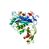
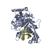
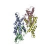
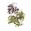
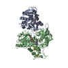
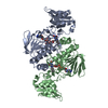
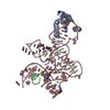
- PDB-1bcm: BACTERIOPHAGE MU TRANSPOSASE CORE DOMAIN WITH 2 MONOMERS PER ASYM... -
+ Open data

- Basic information
| Entry | Database: PDB / ID: 1bcm | ||||||
|---|---|---|---|---|---|---|---|
| Title | BACTERIOPHAGE MU TRANSPOSASE CORE DOMAIN WITH 2 MONOMERS PER ASYMMETRIC UNIT | ||||||
 Components Components | BACTERIOPHAGE MU TRANSPOSASE | ||||||
 Keywords Keywords |  TRANSPOSASE / POLYNUCLEOTIDYL TRANSFERASE / DNA BINDING / ENDONUCLEASE / INTEGRASE TRANSPOSASE / POLYNUCLEOTIDYL TRANSFERASE / DNA BINDING / ENDONUCLEASE / INTEGRASE | ||||||
| Function / homology |  Function and homology informationLigases; Forming phosphoric-ester bonds / Hydrolases; Acting on ester bonds; Endodeoxyribonucleases producing 3'-phosphomonoesters / latency-replication decision / transposase activity / DNA transposition / double-stranded DNA endonuclease activity / viral DNA genome replication / ligase activity / DNA integration / DNA replication ...Ligases; Forming phosphoric-ester bonds / Hydrolases; Acting on ester bonds; Endodeoxyribonucleases producing 3'-phosphomonoesters / latency-replication decision / transposase activity / DNA transposition / double-stranded DNA endonuclease activity / viral DNA genome replication / ligase activity / DNA integration / DNA replication / host cell cytoplasm / DNA repair / DNA binding / metal ion binding Function and homology informationLigases; Forming phosphoric-ester bonds / Hydrolases; Acting on ester bonds; Endodeoxyribonucleases producing 3'-phosphomonoesters / latency-replication decision / transposase activity / DNA transposition / double-stranded DNA endonuclease activity / viral DNA genome replication / ligase activity / DNA integration / DNA replication ...Ligases; Forming phosphoric-ester bonds / Hydrolases; Acting on ester bonds; Endodeoxyribonucleases producing 3'-phosphomonoesters / latency-replication decision / transposase activity / DNA transposition / double-stranded DNA endonuclease activity / viral DNA genome replication / ligase activity / DNA integration / DNA replication / host cell cytoplasm / DNA repair / DNA binding / metal ion bindingSimilarity search - Function | ||||||
| Biological species |  Enterobacteria phage Mu (virus) Enterobacteria phage Mu (virus) | ||||||
| Method | X-RAY DIFFRACTION / Resolution: 2.8 Å | ||||||
 Authors Authors | Rice, P.A. / Mizuuchi, K. | ||||||
 Citation Citation | Journal: Cell(Cambridge,Mass.) / Year: 1995 Title: Structure of the bacteriophage Mu transposase core: a common structural motif for DNA transposition and retroviral integration. Authors: Rice, P. / Mizuuchi, K. | ||||||
| History |
|
- Structure visualization
| Structure viewer | Molecule: MolmilJmol/JSmol |
|---|
- Downloads & links
-Download
| PDBx/mmCIF format | 1bcm.cif.gz | 147.7 KB | Display | PDBx/mmCIF format |
|---|---|---|---|---|
| PDB format | pdb1bcm.ent.gz | 117.2 KB | Display | PDB format |
| PDBx/mmJSON format | 1bcm.json.gz | Tree view | PDBx/mmJSON format | |
| Others |  Other downloads Other downloads |
-Validation report
| Arichive directory | https://data.pdbj.org/pub/pdb/validation_reports/bc/1bcmftp://data.pdbj.org/pub/pdb/validation_reports/bc/1bcm | HTTPS FTP |
|---|
-Related structure data


-Links
 PDBj
PDBj


- Assembly
| Deposited unit | 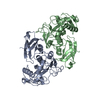
| ||||||||
|---|---|---|---|---|---|---|---|---|---|
| 1 |
| ||||||||
| Unit cell |
| ||||||||
| Noncrystallographic symmetry (NCS) | NCS oper: (Code: given Matrix: (-0.6177, -0.0594, -0.7841), Vector : Details | MTRIX THE TRANSFORMATIONS PRESENTED ON MTRIX RECORDS BELOW DESCRIBE NON-CRYSTALLOGRAPHIC RELATIONSHIPS AMONG THE VARIOUS DOMAINS IN THIS ENTRY. APPLYING THE APPROPRIATE MTRIX TRANSFORMATION TO THE RESIDUES LISTED FIRST WILL YIELD APPROXIMATE COORDINATES FOR THE RESIDUES LISTED SECOND. APPLIED TO TRANSFORMED TO MTRIX RESIDUES RESIDUES RMSD M1 B 258 .. B 560 A 258 .. A 560 0.593 THIS REPRESENTS A NON-CRYSTALLOGRAPHIC TWO-FOLD AXIS. | |
-Components
| #1: Protein | Mass: 36888.078 Da / Num. of mol.: 2 Source method: isolated from a genetically manipulated source Source: (gene. exp.) Enterobacteria phage Mu (virus) / Genus: Mu-like viruses / Strain: WILD TYPE / Gene: MUA (AMINO ACIDS 248 - 574) / Plasmid: PMK602 / Gene (production host): MUA (AMINO ACIDS 248 - 574) / Production host:  Escherichia coli (E. coli) / Strain (production host): T7 / References: UniProt: P07636 Escherichia coli (E. coli) / Strain (production host): T7 / References: UniProt: P07636 |
|---|
-Experimental details
-Experiment
| Experiment | Method: X-RAY DIFFRACTION |
|---|
- Sample preparation
| Crystal | Density Matthews: 2.79 Å3/Da / Density % sol: 55.95 % | ||||||||||||||||||||||||||||||||||||
|---|---|---|---|---|---|---|---|---|---|---|---|---|---|---|---|---|---|---|---|---|---|---|---|---|---|---|---|---|---|---|---|---|---|---|---|---|---|
| Crystal grow | *PLUS Method: vapor diffusion, hanging drop | ||||||||||||||||||||||||||||||||||||
| Components of the solutions | *PLUS
|
-Data collection
| Diffraction source | Wavelength: 1.54 |
|---|---|
| Detector | Type: RIGAKU / Detector: IMAGE PLATE / Date: Aug 27, 1994 |
| Radiation | Monochromatic (M) / Laue (L): M / Scattering type: x-ray |
| Radiation wavelength | Wavelength: 1.54 Å / Relative weight: 1 |
| Reflection | Resolution: 2.7→20 Å / Num. obs: 21089 / % possible obs: 92.5 % / Observed criterion σ(I): 0 / Redundancy: 3.3 % / Rmerge(I) obs: 0.095 |
| Reflection | *PLUS Highest resolution: 2.7 Å / Rmerge(I) obs: 0.095 |
- Processing
| Software |
| ||||||||||||||||||||||||||||||||||||||||||||||||||||||||||||
|---|---|---|---|---|---|---|---|---|---|---|---|---|---|---|---|---|---|---|---|---|---|---|---|---|---|---|---|---|---|---|---|---|---|---|---|---|---|---|---|---|---|---|---|---|---|---|---|---|---|---|---|---|---|---|---|---|---|---|---|---|---|
| Refinement | Resolution: 2.8→8 Å / σ(F): 1 Details: RESIDUES ARG A 355 AND ARG B 355 LIE IN A DISALLOWED REGION OF A RAMACHANDRAN PLOT. THIS WAS CONFIRMED IN THE C 2 2 21 STRUCTURE BY SIMULATED ANNEALING OMIT MAP.
| ||||||||||||||||||||||||||||||||||||||||||||||||||||||||||||
| Displacement parameters | Biso mean: 20.7 Å2 | ||||||||||||||||||||||||||||||||||||||||||||||||||||||||||||
| Refinement step | Cycle: LAST / Resolution: 2.8→8 Å
| ||||||||||||||||||||||||||||||||||||||||||||||||||||||||||||
| Refine LS restraints |
| ||||||||||||||||||||||||||||||||||||||||||||||||||||||||||||
| Software | *PLUS Name: X-PLOR / Version: 3.1 / Classification: refinement | ||||||||||||||||||||||||||||||||||||||||||||||||||||||||||||
| Refine LS restraints | *PLUS
| ||||||||||||||||||||||||||||||||||||||||||||||||||||||||||||
| LS refinement shell | *PLUS Highest resolution: 2.8 Å / Lowest resolution: 2.92 Å |
